 Movie
Movie Controller


+ Open data

- Basic information
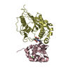
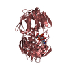
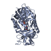
| Entry | Database: PDB / ID: 1lq8 | |||||||||
|---|---|---|---|---|---|---|---|---|---|---|
| Title | Crystal structure of cleaved protein C inhibitor | |||||||||
 Components Components | (plasma serine protease ...) x 2 | |||||||||
 Keywords Keywords | BLOOD CLOTTING / serpin / protease / inhibitor / heparin / retinoic acid / protein C | |||||||||
| Function / homology |  Function and homology information Function and homology informationprotein C inhibitor-TMPRSS7 complex / protein C inhibitor-TMPRSS11E complex / protein C inhibitor-PLAT complex / protein C inhibitor-PLAU complex / protein C inhibitor-thrombin complex / protein C inhibitor-KLK3 complex / protein C inhibitor-plasma kallikrein complex / seminal clot liquefaction / protein C inhibitor-coagulation factor V complex / protein C inhibitor-coagulation factor Xa complex ...protein C inhibitor-TMPRSS7 complex / protein C inhibitor-TMPRSS11E complex / protein C inhibitor-PLAT complex / protein C inhibitor-PLAU complex / protein C inhibitor-thrombin complex / protein C inhibitor-KLK3 complex / protein C inhibitor-plasma kallikrein complex / seminal clot liquefaction / protein C inhibitor-coagulation factor V complex / protein C inhibitor-coagulation factor Xa complex / protein C inhibitor-coagulation factor XI complex / acrosin binding / platelet dense tubular network / fusion of sperm to egg plasma membrane involved in single fertilization / acrosomal membrane / platelet alpha granule / glycosaminoglycan binding / phosphatidylcholine binding / retinoic acid binding / lipid transport / : / : / serine-type endopeptidase inhibitor activity / heparin binding / protease binding / spermatogenesis / external side of plasma membrane / protein-containing complex / : / extracellular exosome / extracellular region / membrane Similarity search - Function | |||||||||
| Biological species |  Homo sapiens (human) Homo sapiens (human) | |||||||||
| Method |  X-RAY DIFFRACTION / SYNCHROTRON / MOLECULAR REPLACEMENT / Resolution: 2.4 Å X-RAY DIFFRACTION / SYNCHROTRON / MOLECULAR REPLACEMENT / Resolution: 2.4 Å | |||||||||
 Authors Authors | Huntington, J.A. / Kjellberg, M. / Stenflo, J. | |||||||||
 Citation Citation | Journal: Structure / Year: 2003 Title: Crystal Structure of Protein C Inhibitor Provides Insights into Hormone Binding and Heparin Activation Authors: Huntington, J.A. / Kjellberg, M. / Stenflo, J. | |||||||||
| History |
|
- Structure visualization
| Structure viewer | Molecule: MolmilJmol/JSmol |
|---|
- Downloads & links
-Download
| PDBx/mmCIF format | 1lq8.cif.gz | 294.1 KB | Display | PDBx/mmCIF format |
|---|---|---|---|---|
| PDB format | pdb1lq8.ent.gz | 238.3 KB | Display | PDB format |
| PDBx/mmJSON format | 1lq8.json.gz | Tree view | PDBx/mmJSON format | |
| Others |  Other downloads Other downloads |
-Validation report
| Arichive directory | https://data.pdbj.org/pub/pdb/validation_reports/lq/1lq8ftp://data.pdbj.org/pub/pdb/validation_reports/lq/1lq8 | HTTPS FTP |
|---|
-Related structure data
| Related structure data |  1ezxS S: Starting model for refinement |
|---|---|
| Similar structure data |


-Links
 PDBj
PDBj


- Assembly
| Deposited unit | 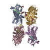
| ||||||||||
|---|---|---|---|---|---|---|---|---|---|---|---|
| 1 | 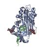
| ||||||||||
| 2 | 
| ||||||||||
| 3 | 
| ||||||||||
| 4 | 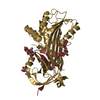
| ||||||||||
| Unit cell |
|
-Components
-Plasma serine protease ... , 2 types, 8 molecules ACEGBDFH
| #1: Protein | Mass: 38651.000 Da / Num. of mol.: 4 Fragment: N-terminal fragment from elastase cleavage, residues 30-375 Source method: isolated from a natural source / Details: Human PCI purified from discarded blood plasma. / Source: (natural) Homo sapiens (human) / Organ: liver / Tissue: blood / Tissue fraction: plasma / References: UniProt: P05154#2: Protein/peptide | Mass: 3737.448 Da / Num. of mol.: 4 Fragment: C-terminal fragment from elastase cleavage, residues 376-405 Source method: isolated from a natural source / Details: Human PCI purified from discarded blood plasma. / Source: (natural) Homo sapiens (human) / Organ: liver / Tissue: blood / Tissue fraction: plasma / References: UniProt: P05154 |
|---|
-Sugars , 4 types, 8 molecules 
| #3: Polysaccharide | Source method: isolated from a genetically manipulated source #4: Polysaccharide | Source method: isolated from a genetically manipulated source #5: Polysaccharide | alpha-D-mannopyranose-(1-3)-[alpha-D-mannopyranose-(1-6)]alpha-D-mannopyranose | Source method: isolated from a genetically manipulated source #7: Sugar |  Type: D-saccharide, beta linking / Mass: 221.208 Da / Num. of mol.: 3 Type: D-saccharide, beta linking / Mass: 221.208 Da / Num. of mol.: 3Source method: isolated from a genetically manipulated source Formula: C8H15NO6 |
|---|
-Non-polymers , 2 types, 102 molecules 

| #6: Chemical |  Mass: 60.095 Da / Num. of mol.: 3 / Source method: obtained synthetically / Formula: C3H8O Mass: 60.095 Da / Num. of mol.: 3 / Source method: obtained synthetically / Formula: C3H8O#8: Water | ChemComp-HOH / | Mass: 18.015 Da / Num. of mol.: 99 / Source method: isolated from a natural source / Formula: H2O |
|---|
-Details
| Has protein modification | Y |
|---|
-Experimental details
-Experiment
| Experiment | Method: X-RAY DIFFRACTION / Number of used crystals: 1 |
|---|
- Sample preparation
| Crystal | Density Matthews: 2.74 Å3/Da / Density % sol: 54.7 % | ||||||||||||||||||||||||||||||
|---|---|---|---|---|---|---|---|---|---|---|---|---|---|---|---|---|---|---|---|---|---|---|---|---|---|---|---|---|---|---|---|
| Crystal grow | Temperature: 293 K / Method: vapor diffusion, hanging drop / pH: 7 Details: PEG 3350, sodium fluoride, isopropanol, pH 7, VAPOR DIFFUSION, HANGING DROP at 293K | ||||||||||||||||||||||||||||||
| Crystal grow | *PLUS Method: vapor diffusion | ||||||||||||||||||||||||||||||
| Components of the solutions | *PLUS
|
-Data collection
| Diffraction | Mean temperature: 100 K |
|---|---|
| Diffraction source | Source: SYNCHROTRON / Site: APS  / Beamline: 19-ID / Wavelength: 1.0332 Å / Beamline: 19-ID / Wavelength: 1.0332 Å |
| Detector | Type: SBC-2 / Detector: CCD / Date: Jan 1, 2002 |
| Radiation | Monochromator: Double crystal: Si-111, Si-220 / Protocol: SINGLE WAVELENGTH / Monochromatic (M) / Laue (L): M / Scattering type: x-ray |
| Radiation wavelength | Wavelength: 1.0332 Å / Relative weight: 1 |
| Reflection | Resolution: 2.4→45.64 Å / Num. all: 68208 / Num. obs: 55034 / % possible obs: 80.7 % / Observed criterion σ(F): 0 / Observed criterion σ(I): 0 / Biso Wilson estimate: 53.85 Å2 / Rmerge(I) obs: 0.148 / Rsym value: 0.112 / Net I/σ(I): 19.2 |
| Reflection shell | Resolution: 2.4→2.49 Å / Rmerge(I) obs: 0.48 / Mean I/σ(I) obs: 1.45 / % possible all: 27.8 |
| Reflection | *PLUS Highest resolution: 2.4 Å / Lowest resolution: 45.6 Å / % possible obs: 81.7 % / Num. measured all: 68208 |
- Processing
| Software |
| |||||||||||||||||||||||||
|---|---|---|---|---|---|---|---|---|---|---|---|---|---|---|---|---|---|---|---|---|---|---|---|---|---|---|
| Refinement | Method to determine structure: MOLECULAR REPLACEMENT Starting model: PDB ENTRY 1EZX Resolution: 2.4→45.66 Å / Isotropic thermal model: anisotropic / Cross valid method: THROUGHOUT / σ(F): 0 / Stereochemistry target values: Engh & Huber / Details: Maximum likelihood
| |||||||||||||||||||||||||
| Displacement parameters |
| |||||||||||||||||||||||||
| Refine analyze |
| |||||||||||||||||||||||||
| Refinement step | Cycle: LAST / Resolution: 2.4→45.66 Å
| |||||||||||||||||||||||||
| Refine LS restraints |
| |||||||||||||||||||||||||
| Refinement | *PLUS Highest resolution: 2.4 Å / Lowest resolution: 45.6 Å | |||||||||||||||||||||||||
| Solvent computation | *PLUS | |||||||||||||||||||||||||
| Displacement parameters | *PLUS |
