 Movie
Movie Controller


+ Open data

- Basic information
| Entry | Database: PDB / ID: 6ndq | ||||||
|---|---|---|---|---|---|---|---|
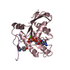
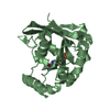
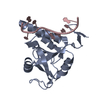
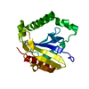
| Title | Crystal structure of a LPMO from Kitasatospora papulosa | ||||||
 Components Components | lytic polysaccharide monooxygenase | ||||||
 Keywords Keywords | OXIDOREDUCTASE / Lytic polysaccharide monooxygenase | ||||||
| Function / homology | chitin-binding protein cbp21 / Cellulose/chitin-binding protein, N-terminal / Lytic polysaccharide mono-oxygenase, cellulose-degrading / Coagulation Factor XIII; Chain A, domain 1 / Distorted Sandwich / Immunoglobulin E-set / Mainly Beta / Chitin-binding domain 3 protein Function and homology information Function and homology information | ||||||
| Biological species |  [Kitasatospora] papulosa (bacteria) [Kitasatospora] papulosa (bacteria) | ||||||
| Method |  X-RAY DIFFRACTION / SYNCHROTRON / MOLECULAR REPLACEMENT / Resolution: 1.601 Å X-RAY DIFFRACTION / SYNCHROTRON / MOLECULAR REPLACEMENT / Resolution: 1.601 Å | ||||||
 Authors Authors | Correa, T.L.C. / Tomazini Jr., A. / Murakami, M.T. | ||||||
| Funding support |  Brazil, 1items Brazil, 1items
| ||||||
 Citation Citation | Journal: To Be Published Title: Crystal structure of a LPMO from Kitasatospora papulosa Authors: Correa, T.L.C. / Tomazini Jr., A. / Murakami, M.T. | ||||||
| History |
|
- Structure visualization
| Structure viewer | Molecule: MolmilJmol/JSmol |
|---|
- Downloads & links
-Download
| PDBx/mmCIF format | 6ndq.cif.gz | 91.1 KB | Display | PDBx/mmCIF format |
|---|---|---|---|---|
| PDB format | pdb6ndq.ent.gz | 68.1 KB | Display | PDB format |
| PDBx/mmJSON format | 6ndq.json.gz | Tree view | PDBx/mmJSON format | |
| Others |  Other downloads Other downloads |
-Validation report
| Arichive directory | https://data.pdbj.org/pub/pdb/validation_reports/nd/6ndqftp://data.pdbj.org/pub/pdb/validation_reports/nd/6ndq | HTTPS FTP |
|---|
-Related structure data
| Related structure data |  4oy6S S: Starting model for refinement |
|---|---|
| Similar structure data |


-Links
 PDBj
PDBj- Assembly
| Deposited unit | 
| ||||||||
|---|---|---|---|---|---|---|---|---|---|
| 1 | 
| ||||||||
| 2 | 
| ||||||||
| Unit cell |
|
-Components
| #1: Protein | Mass: 21662.461 Da / Num. of mol.: 2 Source method: isolated from a genetically manipulated source Source: (gene. exp.) [Kitasatospora] papulosa (bacteria) / Production host: #2: Water | ChemComp-HOH / |  Mass: 18.015 Da / Num. of mol.: 212 / Source method: isolated from a natural source / Formula: H2O Mass: 18.015 Da / Num. of mol.: 212 / Source method: isolated from a natural source / Formula: H2OHas protein modification | Y | |
|---|
-Experimental details
-Experiment
| Experiment | Method: X-RAY DIFFRACTION / Number of used crystals: 1 |
|---|
- Sample preparation
| Crystal | Density Matthews: 1.85 Å3/Da / Density % sol: 33.38 % |
|---|---|
| Crystal grow | Temperature: 291 K / Method: vapor diffusion, sitting drop / Details: 0.1 M MMT buffer pH 4.0 25% PEG1500 |
-Data collection
| Diffraction | Mean temperature: 100 K / Serial crystal experiment: N |
|---|---|
| Diffraction source | Source: SYNCHROTRON / Site: LNLS / Beamline: W01B-MX2 / Wavelength: 1.453 Å |
| Detector | Type: DECTRIS PILATUS 2M / Detector: PIXEL / Date: Sep 6, 2018 |
| Radiation | Protocol: SINGLE WAVELENGTH / Monochromatic (M) / Laue (L): M / Scattering type: x-ray |
| Radiation wavelength | Wavelength: 1.453 Å / Relative weight: 1 |
| Reflection | Resolution: 1.6→39.24 Å / Num. obs: 39856 / % possible obs: 96.3 % / Redundancy: 3.18 % / Net I/σ(I): 8.9 |
| Reflection shell | Resolution: 1.6→1.7 Å |
- Processing
| Software |
| |||||||||||||||||||||||||||||||||||||||||||||||||||||||||||||||||||||||||||||||||||||||||||||||||||||||||
|---|---|---|---|---|---|---|---|---|---|---|---|---|---|---|---|---|---|---|---|---|---|---|---|---|---|---|---|---|---|---|---|---|---|---|---|---|---|---|---|---|---|---|---|---|---|---|---|---|---|---|---|---|---|---|---|---|---|---|---|---|---|---|---|---|---|---|---|---|---|---|---|---|---|---|---|---|---|---|---|---|---|---|---|---|---|---|---|---|---|---|---|---|---|---|---|---|---|---|---|---|---|---|---|---|---|---|
| Refinement | Method to determine structure: MOLECULAR REPLACEMENT Starting model: 4OY6 Resolution: 1.601→33.088 Å / SU ML: 0.23 / Cross valid method: FREE R-VALUE / σ(F): 0 / Phase error: 27.97
| |||||||||||||||||||||||||||||||||||||||||||||||||||||||||||||||||||||||||||||||||||||||||||||||||||||||||
| Solvent computation | Shrinkage radii: 0.9 Å / VDW probe radii: 1.11 Å | |||||||||||||||||||||||||||||||||||||||||||||||||||||||||||||||||||||||||||||||||||||||||||||||||||||||||
| Refinement step | Cycle: LAST / Resolution: 1.601→33.088 Å
| |||||||||||||||||||||||||||||||||||||||||||||||||||||||||||||||||||||||||||||||||||||||||||||||||||||||||
| Refine LS restraints |
| |||||||||||||||||||||||||||||||||||||||||||||||||||||||||||||||||||||||||||||||||||||||||||||||||||||||||
| LS refinement shell |
|
