- PDB-1z6x: Structure Of Human ADP-Ribosylation Factor 4 -
+
Open data
ID or keywords:
Loading...
-
Basic information
Entry
Database: PDB / ID: 1z6x
Title
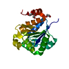
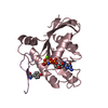
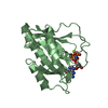
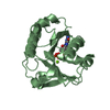
Structure Of Human ADP-Ribosylation Factor 4
Components
ADP-ribosylation factor 4
Keywords
TRANSPORT PROTEIN / GDP-BINDING / MEMBRANE TRAFFICKING / GOLGI STACK / LIPOPROTEIN / MYRISTATE / Structural Genomics / PSI / Protein Structure Initiative / Structural Genomics Consortium / SGC
Function / homology
Function and homology information
phospholipase D activator activity / regulation of cilium assembly / NAD+-protein-arginine ADP-ribosyltransferase activity / VxPx cargo-targeting to cilium / dendritic spine development / establishment or maintenance of epithelial cell apical/basal polarity / protein localization to cilium / retrograde vesicle-mediated transport, Golgi to endoplasmic reticulum / apical protein localization / regulation of postsynapse organization ...phospholipase D activator activity / regulation of cilium assembly / NAD+-protein-arginine ADP-ribosyltransferase activity / VxPx cargo-targeting to cilium / dendritic spine development / establishment or maintenance of epithelial cell apical/basal polarity / protein localization to cilium / retrograde vesicle-mediated transport, Golgi to endoplasmic reticulum / apical protein localization / regulation of postsynapse organization / epidermal growth factor receptor binding / regulation of reactive oxygen species metabolic process / COPI-dependent Golgi-to-ER retrograde traffic / cilium assembly / endoplasmic reticulum to Golgi vesicle-mediated transport / response to axon injury / COPI-mediated anterograde transport / vesicle-mediated transport / guanyl-nucleotide exchange factor activity / learning / intracellular protein transport / epidermal growth factor receptor signaling pathway / ruffle membrane / cell migration / dendritic spine / Golgi membrane / GTPase activity / negative regulation of apoptotic process / GTP binding / glutamatergic synapse / positive regulation of transcription by RNA polymerase II / extracellular exosome / membrane / plasma membrane / cytoplasm / cytosol Similarity search - Function
ADP-ribosylation factor 1-5 / Small GTPase superfamily, ARF type / Small GTPase Arf domain profile. / Sar1p-like members of the Ras-family of small GTPases / Small GTPase superfamily, ARF/SAR type / ADP-ribosylation factor family / ARF-like small GTPases; ARF, ADP-ribosylation factor / Rab subfamily of small GTPases / Small GTP-binding protein domain / P-loop containing nucleotide triphosphate hydrolases ...ADP-ribosylation factor 1-5 / Small GTPase superfamily, ARF type / Small GTPase Arf domain profile. / Sar1p-like members of the Ras-family of small GTPases / Small GTPase superfamily, ARF/SAR type / ADP-ribosylation factor family / ARF-like small GTPases; ARF, ADP-ribosylation factor / Rab subfamily of small GTPases / Small GTP-binding protein domain / P-loop containing nucleotide triphosphate hydrolases / Rossmann fold / P-loop containing nucleoside triphosphate hydrolase / 3-Layer(aba) Sandwich / Alpha Beta Similarity search - Domain/homology
Resolution: 2.7→32.38 Å / Cor.coef. Fo:Fc: 0.918 / Cor.coef. Fo:Fc free: 0.882 / Cross valid method: THROUGHOUT / ESU R Free: 0.422 / Stereochemistry target values: MAXIMUM LIKELIHOOD / Details: HYDROGENS HAVE BEEN ADDED IN THE RIDING POSITIONS
Rfactor
Num. reflection
% reflection
Selection details
Rfree
0.27269
466
4.8 %
RANDOM
Rwork
0.22166
-
-
-
obs
0.22419
9184
99.84 %
-
all
-
9651
-
-
Solvent computation
Ion probe radii: 0.8 Å / Shrinkage radii: 0.8 Å / VDW probe radii: 1.2 Å / Solvent model: MASK
Displacement parameters
Biso mean: 40.803 Å2
Baniso -1
Baniso -2
Baniso -3
1-
1.18 Å2
0 Å2
0 Å2
2-
-
1.18 Å2
0 Å2
3-
-
-
-2.37 Å2
Refinement step
Cycle: LAST / Resolution: 2.7→32.38 Å
Protein
Nucleic acid
Ligand
Solvent
Total
Num. atoms
2800
0
58
55
2913
Refine LS restraints
Refine-ID
Type
Dev ideal
Dev ideal target
Number
X-RAY DIFFRACTION
r_bond_refined_d
0.009
0.022
2906
X-RAY DIFFRACTION
r_bond_other_d
X-RAY DIFFRACTION
r_angle_refined_deg
1.226
1.988
3944
X-RAY DIFFRACTION
r_angle_other_deg
X-RAY DIFFRACTION
r_dihedral_angle_1_deg
5.124
5
348
X-RAY DIFFRACTION
r_dihedral_angle_2_deg
43.67
24.615
130
X-RAY DIFFRACTION
r_dihedral_angle_3_deg
15.955
15
528
X-RAY DIFFRACTION
r_dihedral_angle_4_deg
18.82
15
18
X-RAY DIFFRACTION
r_chiral_restr
0.07
0.2
456
X-RAY DIFFRACTION
r_gen_planes_refined
0.003
0.02
2112
X-RAY DIFFRACTION
r_gen_planes_other
X-RAY DIFFRACTION
r_nbd_refined
0.23
0.2
1519
X-RAY DIFFRACTION
r_nbd_other
X-RAY DIFFRACTION
r_nbtor_refined
0.311
0.2
1988
X-RAY DIFFRACTION
r_nbtor_other
X-RAY DIFFRACTION
r_xyhbond_nbd_refined
0.161
0.2
141
X-RAY DIFFRACTION
r_xyhbond_nbd_other
X-RAY DIFFRACTION
r_metal_ion_refined
X-RAY DIFFRACTION
r_metal_ion_other
X-RAY DIFFRACTION
r_symmetry_vdw_refined
0.236
0.2
75
X-RAY DIFFRACTION
r_symmetry_vdw_other
X-RAY DIFFRACTION
r_symmetry_hbond_refined
0.291
0.2
6
X-RAY DIFFRACTION
r_symmetry_hbond_other
X-RAY DIFFRACTION
r_symmetry_metal_ion_refined
X-RAY DIFFRACTION
r_symmetry_metal_ion_other
X-RAY DIFFRACTION
r_mcbond_it
0.605
1.5
1730
X-RAY DIFFRACTION
r_mcbond_other
X-RAY DIFFRACTION
r_mcangle_it
1.102
2
2796
X-RAY DIFFRACTION
r_scbond_it
0.799
3
1176
X-RAY DIFFRACTION
r_scangle_it
1.348
4.5
1148
X-RAY DIFFRACTION
r_rigid_bond_restr
X-RAY DIFFRACTION
r_sphericity_free
X-RAY DIFFRACTION
r_sphericity_bonded
LS refinement shell
Resolution: 2.7→2.77 Å / Total num. of bins used: 20
Rfactor
Num. reflection
% reflection
Rfree
0.385
27
-
Rwork
0.295
642
-
obs
-
-
100 %
+
About Yorodumi
-
News
-
Feb 9, 2022. New format data for meta-information of EMDB entries
New format data for meta-information of EMDB entries
Version 3 of the EMDB header file is now the official format.
The previous official version 1.9 will be removed from the archive.
In the structure databanks used in Yorodumi, some data are registered as the other names, "COVID-19 virus" and "2019-nCoV". Here are the details of the virus and the list of structure data.
Jan 31, 2019. EMDB accession codes are about to change! (news from PDBe EMDB page)
EMDB accession codes are about to change! (news from PDBe EMDB page)
The allocation of 4 digits for EMDB accession codes will soon come to an end. Whilst these codes will remain in use, new EMDB accession codes will include an additional digit and will expand incrementally as the available range of codes is exhausted. The current 4-digit format prefixed with “EMD-” (i.e. EMD-XXXX) will advance to a 5-digit format (i.e. EMD-XXXXX), and so on. It is currently estimated that the 4-digit codes will be depleted around Spring 2019, at which point the 5-digit format will come into force.
The EM Navigator/Yorodumi systems omit the EMD- prefix.
Related info.:Q: What is EMD? / ID/Accession-code notation in Yorodumi/EM Navigator
Yorodumi is a browser for structure data from EMDB, PDB, SASBDB, etc.
This page is also the successor to EM Navigator detail page, and also detail information page/front-end page for Omokage search.
The word "yorodu" (or yorozu) is an old Japanese word meaning "ten thousand". "mi" (miru) is to see.
Related info.:EMDB / PDB / SASBDB / Comparison of 3 databanks / Yorodumi Search / Aug 31, 2016. New EM Navigator & Yorodumi / Yorodumi Papers / Jmol/JSmol / Function and homology information / Changes in new EM Navigator and Yorodumi
 Movie
Movie Controller


 Open data
 Basic information
 Components
 Keywords
 Function and homology information
 Homo sapiens (human)
 X-RAY DIFFRACTION /
  Authors
 Citation
 Structure visualization
 Downloads & links
 Other downloads
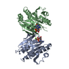


 PDBj
PDBj


 Assembly


 Mass: 24.305 Da / Num. of mol.: 2 / Source method: obtained synthetically / Formula: Mg
 Type: RNA linking / Mass: 443.201 Da / Num. of mol.: 2 / Source method: obtained synthetically / Formula: C10H15N5O11P2 / Comment: GDP, energy-carrying molecule*YM
 Mass: 18.015 Da / Num. of mol.: 55 / Source method: isolated from a natural source / Formula: H2O
 Sample preparation
 Processing