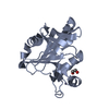
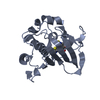
 Movie
Movie Controller


+ Open data

- Basic information
| Entry | Database: PDB / ID: 3rzu | ||||||
|---|---|---|---|---|---|---|---|
| Title | The Crystal Structure of the Catalytic Domain of AMSH | ||||||
 Components Components | STAM-binding protein | ||||||
 Keywords Keywords | HYDROLASE / Ubiquitin Hydrolase / STAM / Endosome-associated deubiquitinating enzyme | ||||||
| Function / homology |  Function and homology information Function and homology informationHydrolases; Acting on peptide bonds (peptidases); Omega peptidases / negative regulation of hippocampal neuron apoptotic process / deubiquitinase activity / negative regulation of Ras protein signal transduction / hippocampal neuron apoptotic process / protein K63-linked deubiquitination / metal-dependent deubiquitinase activity / K63-linked deubiquitinase activity / cleavage furrow / mitotic cytokinesis ...Hydrolases; Acting on peptide bonds (peptidases); Omega peptidases / negative regulation of hippocampal neuron apoptotic process / deubiquitinase activity / negative regulation of Ras protein signal transduction / hippocampal neuron apoptotic process / protein K63-linked deubiquitination / metal-dependent deubiquitinase activity / K63-linked deubiquitinase activity / cleavage furrow / mitotic cytokinesis / protein deubiquitination / cell surface receptor signaling pathway via JAK-STAT / negative regulation of phosphatidylinositol 3-kinase/protein kinase B signal transduction / Metalloprotease DUBs / early endosome / endosome / protein domain specific binding / positive regulation of cell population proliferation / proteolysis / extracellular exosome / nucleoplasm / metal ion binding / nucleus / plasma membrane / cytosol Similarity search - Function | ||||||
| Biological species |  Homo sapiens (human) Homo sapiens (human) | ||||||
| Method |  X-RAY DIFFRACTION / SYNCHROTRON / MOLECULAR REPLACEMENT / Resolution: 2.5 Å X-RAY DIFFRACTION / SYNCHROTRON / MOLECULAR REPLACEMENT / Resolution: 2.5 Å | ||||||
 Authors Authors | Davies, C.W. / Das, C. | ||||||
 Citation Citation | Journal: J.Mol.Biol. / Year: 2011 Title: Structural and Thermodynamic Comparison of the Catalytic Domain of AMSH and AMSH-LP: Nearly Identical Fold but Different Stability. Authors: Davies, C.W. / Paul, L.N. / Kim, M.I. / Das, C. | ||||||
| History |
|
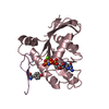
- Structure visualization
| Structure viewer | Molecule: MolmilJmol/JSmol |
|---|
- Downloads & links
-Download
| PDBx/mmCIF format | 3rzu.cif.gz | 484.9 KB | Display | PDBx/mmCIF format |
|---|---|---|---|---|
| PDB format | pdb3rzu.ent.gz | 401.3 KB | Display | PDB format |
| PDBx/mmJSON format | 3rzu.json.gz | Tree view | PDBx/mmJSON format | |
| Others |  Other downloads Other downloads |
-Validation report
| Arichive directory | https://data.pdbj.org/pub/pdb/validation_reports/rz/3rzuftp://data.pdbj.org/pub/pdb/validation_reports/rz/3rzu | HTTPS FTP |
|---|
-Related structure data
| Related structure data |  3rzvC  2znrS S: Starting model for refinement C: citing same article ( |
|---|---|
| Similar structure data |


-Links
 PDBj
PDBj

- Assembly
| Deposited unit | 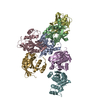
| ||||||||
|---|---|---|---|---|---|---|---|---|---|
| 1 | 
| ||||||||
| 2 | 
| ||||||||
| 3 | 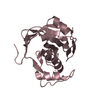
| ||||||||
| 4 | 
| ||||||||
| 5 | 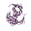
| ||||||||
| 6 | 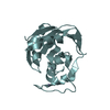
| ||||||||
| 7 | 
| ||||||||
| Unit cell |
|
-Components
| #1: Protein | Mass: 20616.455 Da / Num. of mol.: 7 / Fragment: Catalytic Domain, residues 243-424 Source method: isolated from a genetically manipulated source Source: (gene. exp.) Homo sapiens (human) / Gene: AMSH, STAMBP, STAMBP (AMSH) / Plasmid: pGEX-6P1 / Production host:  References: UniProt: O95630, Hydrolases; Acting on peptide bonds (peptidases); Omega peptidases #2: Chemical | ChemComp-ZN /   Mass: 65.409 Da / Num. of mol.: 14 / Source method: obtained synthetically / Formula: Zn Mass: 65.409 Da / Num. of mol.: 14 / Source method: obtained synthetically / Formula: Zn#3: Water | ChemComp-HOH / |  Mass: 18.015 Da / Num. of mol.: 173 / Source method: isolated from a natural source / Formula: H2O Mass: 18.015 Da / Num. of mol.: 173 / Source method: isolated from a natural source / Formula: H2O |
|---|
-Experimental details
-Experiment
| Experiment | Method: X-RAY DIFFRACTION / Number of used crystals: 1 |
|---|
- Sample preparation
| Crystal | Density Matthews: 2.48 Å3/Da / Density % sol: 50.48 % |
|---|---|
| Crystal grow | Temperature: 298 K / Method: vapor diffusion, sitting drop / pH: 7 Details: 0.2M Sodium malonate, 20% PEG 3350, 5% PEG 400, pH 7.0, VAPOR DIFFUSION, SITTING DROP, temperature 298K |
-Data collection
| Diffraction | Mean temperature: 100 K | |||||||||||||||||||||||||||||||||||||||||||||||||||||||
|---|---|---|---|---|---|---|---|---|---|---|---|---|---|---|---|---|---|---|---|---|---|---|---|---|---|---|---|---|---|---|---|---|---|---|---|---|---|---|---|---|---|---|---|---|---|---|---|---|---|---|---|---|---|---|---|---|
| Diffraction source | Source: SYNCHROTRON / Site: APS  / Beamline: 23-ID-B / Wavelength: 1 Å / Beamline: 23-ID-B / Wavelength: 1 Å | |||||||||||||||||||||||||||||||||||||||||||||||||||||||
| Detector | Type: MARMOSAIC 300 mm CCD / Detector: CCD / Date: Jul 10, 2010 / Details: Mirrors | |||||||||||||||||||||||||||||||||||||||||||||||||||||||
| Radiation | Monochromator: Si 111 / Protocol: SINGLE WAVELENGTH / Monochromatic (M) / Laue (L): M / Scattering type: x-ray | |||||||||||||||||||||||||||||||||||||||||||||||||||||||
| Radiation wavelength | Wavelength: 1 Å / Relative weight: 1 | |||||||||||||||||||||||||||||||||||||||||||||||||||||||
| Reflection | Resolution: 2.5→50 Å / Num. all: 49199 / Num. obs: 48953 / % possible obs: 99.5 % / Observed criterion σ(F): 2 / Observed criterion σ(I): 2 / Redundancy: 3.5 % / Biso Wilson estimate: 47.5 Å2 / Rsym value: 0.085 / Net I/σ(I): 12.6 | |||||||||||||||||||||||||||||||||||||||||||||||||||||||
| Reflection shell |
|
- Processing
| Software |
| |||||||||||||||||||||||||||||||||||||||||||||||||||||||||||||||||||||||||||||||||||||||||||||||||||||||||||||||||||||||||||||||||||||
|---|---|---|---|---|---|---|---|---|---|---|---|---|---|---|---|---|---|---|---|---|---|---|---|---|---|---|---|---|---|---|---|---|---|---|---|---|---|---|---|---|---|---|---|---|---|---|---|---|---|---|---|---|---|---|---|---|---|---|---|---|---|---|---|---|---|---|---|---|---|---|---|---|---|---|---|---|---|---|---|---|---|---|---|---|---|---|---|---|---|---|---|---|---|---|---|---|---|---|---|---|---|---|---|---|---|---|---|---|---|---|---|---|---|---|---|---|---|---|---|---|---|---|---|---|---|---|---|---|---|---|---|---|---|---|
| Refinement | Method to determine structure: MOLECULAR REPLACEMENT Starting model: 2ZNR Resolution: 2.5→47.152 Å / SU ML: 0.57 / σ(F): 1.34 / Phase error: 24.4 / Stereochemistry target values: ML
| |||||||||||||||||||||||||||||||||||||||||||||||||||||||||||||||||||||||||||||||||||||||||||||||||||||||||||||||||||||||||||||||||||||
| Solvent computation | Shrinkage radii: 0.73 Å / VDW probe radii: 1 Å / Solvent model: FLAT BULK SOLVENT MODEL / Bsol: 51.664 Å2 / ksol: 0.334 e/Å3 | |||||||||||||||||||||||||||||||||||||||||||||||||||||||||||||||||||||||||||||||||||||||||||||||||||||||||||||||||||||||||||||||||||||
| Displacement parameters |
| |||||||||||||||||||||||||||||||||||||||||||||||||||||||||||||||||||||||||||||||||||||||||||||||||||||||||||||||||||||||||||||||||||||
| Refinement step | Cycle: LAST / Resolution: 2.5→47.152 Å
| |||||||||||||||||||||||||||||||||||||||||||||||||||||||||||||||||||||||||||||||||||||||||||||||||||||||||||||||||||||||||||||||||||||
| Refine LS restraints |
| |||||||||||||||||||||||||||||||||||||||||||||||||||||||||||||||||||||||||||||||||||||||||||||||||||||||||||||||||||||||||||||||||||||
| LS refinement shell |
| |||||||||||||||||||||||||||||||||||||||||||||||||||||||||||||||||||||||||||||||||||||||||||||||||||||||||||||||||||||||||||||||||||||
| Refinement TLS params. | Method: refined / Origin x: 33.5007 Å / Origin y: 9.6836 Å / Origin z: 18.7944 Å
| |||||||||||||||||||||||||||||||||||||||||||||||||||||||||||||||||||||||||||||||||||||||||||||||||||||||||||||||||||||||||||||||||||||
| Refinement TLS group | Selection details: all |
