 Movie
Movie Controller


+ Open data

- Basic information
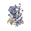
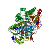
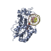
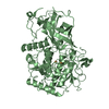
| Entry | Database: PDB / ID: 7mht | ||||||
|---|---|---|---|---|---|---|---|
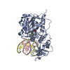
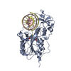
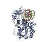
| Title | CYTOSINE-SPECIFIC METHYLTRANSFERASE HHAI/DNA COMPLEX | ||||||
 Components Components |
| ||||||
 Keywords Keywords | TRANSFERASE/DNA / TRANSFERASE / METHYLTRANSFERASE / RESTRICTION SYSTEM / COMPLEX (METHYLTRANSFERASE- DNA) / TRANSFERASE-DNA COMPLEX | ||||||
| Function / homology |  Function and homology information Function and homology informationDNA (cytosine-5-)-methyltransferase / DNA (cytosine-5-)-methyltransferase activity / DNA restriction-modification system / methylation / DNA binding Similarity search - Function | ||||||
| Biological species |  Haemophilus haemolyticus (bacteria) Haemophilus haemolyticus (bacteria) | ||||||
| Method |  X-RAY DIFFRACTION / SYNCHROTRON / OTHER / Resolution: 2.87 Å X-RAY DIFFRACTION / SYNCHROTRON / OTHER / Resolution: 2.87 Å | ||||||
 Authors Authors | O'Gara, M. / Horton, J.R. / Roberts, R.J. / Cheng, X. | ||||||
 Citation Citation | Journal: Nat.Struct.Biol. / Year: 1998 Title: Structures of HhaI methyltransferase complexed with substrates containing mismatches at the target base. Authors: O'Gara, M. / Horton, J.R. / Roberts, R.J. / Cheng, X. | ||||||
| History |
|
- Structure visualization
| Structure viewer | Molecule: MolmilJmol/JSmol |
|---|
- Downloads & links
-Download
| PDBx/mmCIF format | 7mht.cif.gz | 97.3 KB | Display | PDBx/mmCIF format |
|---|---|---|---|---|
| PDB format | pdb7mht.ent.gz | 70.1 KB | Display | PDB format |
| PDBx/mmJSON format | 7mht.json.gz | Tree view | PDBx/mmJSON format | |
| Others |  Other downloads Other downloads |
-Validation report
| Arichive directory | https://data.pdbj.org/pub/pdb/validation_reports/mh/7mhtftp://data.pdbj.org/pub/pdb/validation_reports/mh/7mht | HTTPS FTP |
|---|
-Related structure data
| Related structure data |  8mhtC  9mhtC 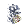 5mhtS S: Starting model for refinement C: citing same article ( |
|---|---|
| Similar structure data |


-Links
 PDBj
PDBj


- Assembly
| Deposited unit | 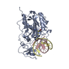
| ||||||||||
|---|---|---|---|---|---|---|---|---|---|---|---|
| 1 |
| ||||||||||
| Unit cell |
|
-Components
| #1: DNA chain | Mass: 3703.416 Da / Num. of mol.: 1 / Source method: obtained synthetically |
|---|---|
| #2: DNA chain | Mass: 3647.393 Da / Num. of mol.: 1 / Source method: obtained synthetically |
| #3: Protein | Mass: 37042.207 Da / Num. of mol.: 1 Source method: isolated from a genetically manipulated source Source: (gene. exp.) Haemophilus haemolyticus (bacteria) / Production host: |
| #4: Chemical | ChemComp-SAH /   Type: L-peptide linking / Mass: 384.411 Da / Num. of mol.: 1 / Source method: obtained synthetically / Formula: C14H20N6O5S Type: L-peptide linking / Mass: 384.411 Da / Num. of mol.: 1 / Source method: obtained synthetically / Formula: C14H20N6O5S |
| #5: Water | ChemComp-HOH /  Mass: 18.015 Da / Num. of mol.: 139 / Source method: isolated from a natural source / Formula: H2O Mass: 18.015 Da / Num. of mol.: 139 / Source method: isolated from a natural source / Formula: H2O |
-Experimental details
-Experiment
| Experiment | Method: X-RAY DIFFRACTION / Number of used crystals: 1 |
|---|
- Sample preparation
| Crystal | Density Matthews: 3.51 Å3/Da / Density % sol: 65 % | ||||||||||||||||||||||||||||||||||||||||||||||||||||||||||||
|---|---|---|---|---|---|---|---|---|---|---|---|---|---|---|---|---|---|---|---|---|---|---|---|---|---|---|---|---|---|---|---|---|---|---|---|---|---|---|---|---|---|---|---|---|---|---|---|---|---|---|---|---|---|---|---|---|---|---|---|---|---|
| Crystal grow | pH: 5.6 / Details: 1.4-1.8 M AMMONIUM SULFATE 50 MM CITRATE PH 5.6 | ||||||||||||||||||||||||||||||||||||||||||||||||||||||||||||
| Components of the solutions |
| ||||||||||||||||||||||||||||||||||||||||||||||||||||||||||||
| Crystal grow | *PLUS Temperature: 16 ℃ / pH: 7.25 / Method: unknown | ||||||||||||||||||||||||||||||||||||||||||||||||||||||||||||
| Components of the solutions | *PLUS
|
-Data collection
| Diffraction | Mean temperature: 293 K |
|---|---|
| Diffraction source | Source: SYNCHROTRON / Site: NSLS  / Beamline: X12C / Wavelength: 1.15 / Beamline: X12C / Wavelength: 1.15 |
| Detector | Type: MARRESEARCH / Detector: IMAGE PLATE |
| Radiation | Protocol: SINGLE WAVELENGTH / Monochromatic (M) / Laue (L): M / Scattering type: x-ray |
| Radiation wavelength | Wavelength: 1.15 Å / Relative weight: 1 |
| Reflection | Resolution: 2.87→23 Å / Num. obs: 13031 / % possible obs: 90 % / Redundancy: 4.3 % / Rmerge(I) obs: 0.066 / Net I/σ(I): 13.3 |
| Reflection | *PLUS % possible obs: 90 % / Observed criterion σ(I): 2 / Num. measured all: 55718 |
- Processing
| Software |
| ||||||||||||||||||||||||||||||||||||||||||||||||||||||||||||
|---|---|---|---|---|---|---|---|---|---|---|---|---|---|---|---|---|---|---|---|---|---|---|---|---|---|---|---|---|---|---|---|---|---|---|---|---|---|---|---|---|---|---|---|---|---|---|---|---|---|---|---|---|---|---|---|---|---|---|---|---|---|
| Refinement | Method to determine structure: OTHER Starting model: PDB ENTRY 5MHT Resolution: 2.87→23 Å / σ(F): 0
| ||||||||||||||||||||||||||||||||||||||||||||||||||||||||||||
| Refinement step | Cycle: LAST / Resolution: 2.87→23 Å
| ||||||||||||||||||||||||||||||||||||||||||||||||||||||||||||
| Refine LS restraints |
| ||||||||||||||||||||||||||||||||||||||||||||||||||||||||||||
| LS refinement shell | Resolution: 2.87→3 Å / Total num. of bins used: 8
| ||||||||||||||||||||||||||||||||||||||||||||||||||||||||||||
| Software | *PLUS Name: X-PLOR / Version: 3.1 / Classification: refinement | ||||||||||||||||||||||||||||||||||||||||||||||||||||||||||||
| Refinement | *PLUS Lowest resolution: 23 Å / σ(F): 0 | ||||||||||||||||||||||||||||||||||||||||||||||||||||||||||||
| Solvent computation | *PLUS | ||||||||||||||||||||||||||||||||||||||||||||||||||||||||||||
| Displacement parameters | *PLUS | ||||||||||||||||||||||||||||||||||||||||||||||||||||||||||||
| Refine LS restraints | *PLUS
| ||||||||||||||||||||||||||||||||||||||||||||||||||||||||||||
| LS refinement shell | *PLUS Rfactor Rwork: 0.199 / Rfactor obs: 0.199 |
