 Movie
Movie Controller


+ Open data

- Basic information
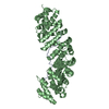
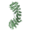
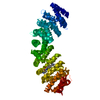
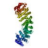
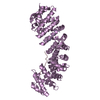
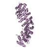
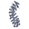
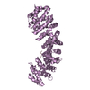
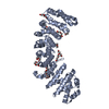
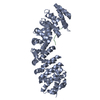
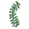
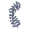
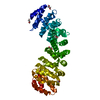
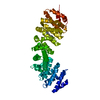
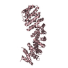
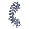
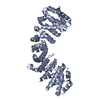
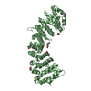
| Entry | Database: PDB / ID: 1q1t | ||||||
|---|---|---|---|---|---|---|---|
| Title | Mouse Importin alpha: non-phosphorylated SV40 CN peptide complex | ||||||
 Components Components |
| ||||||
 Keywords Keywords | PROTEIN TRANSPORT / importin alpha/karyopherin alpha / nuclear localisation sequence (NLS) recognition / phosphorylation / simian virus (SV40) large tumor-antigen (T-antigen) NLS / X-ray crystal structure | ||||||
| Function / homology |  Function and homology information Function and homology informationsymbiont-mediated suppression of host JAK-STAT cascade via inhibition of JAK1 activity / Sensing of DNA Double Strand Breaks / regulation of transcription by glucose / entry of viral genome into host nucleus through nuclear pore complex via importin / positive regulation of viral life cycle / NLS-dependent protein nuclear import complex / postsynapse to nucleus signaling pathway / bidirectional double-stranded viral DNA replication / viral DNA genome replication / nuclear import signal receptor activity ...symbiont-mediated suppression of host JAK-STAT cascade via inhibition of JAK1 activity / Sensing of DNA Double Strand Breaks / regulation of transcription by glucose / entry of viral genome into host nucleus through nuclear pore complex via importin / positive regulation of viral life cycle / NLS-dependent protein nuclear import complex / postsynapse to nucleus signaling pathway / bidirectional double-stranded viral DNA replication / viral DNA genome replication / nuclear import signal receptor activity / nuclear localization sequence binding / NLS-bearing protein import into nucleus / non-canonical NF-kappaB signal transduction / symbiont-mediated perturbation of host cell cycle G1/S transition checkpoint / 3'-5' DNA helicase activity / DNA 3'-5' helicase / DNA replication origin binding / positive regulation of type I interferon production / protein import into nucleus / histone deacetylase binding / cytoplasmic stress granule / host cell / single-stranded DNA binding / double-stranded DNA binding / nuclear membrane / DNA-binding transcription factor binding / symbiont-mediated perturbation of host ubiquitin-like protein modification / DNA replication / postsynaptic density / symbiont-mediated suppression of host innate immune response / symbiont-mediated suppression of host type I interferon-mediated signaling pathway / positive regulation of DNA-templated transcription / host cell nucleus / glutamatergic synapse / DNA-templated transcription / ATP hydrolysis activity / zinc ion binding / nucleoplasm / ATP binding / identical protein binding / nucleus / cytosol / cytoplasm Similarity search - Function | ||||||
| Biological species |  | ||||||
| Method |  X-RAY DIFFRACTION / SYNCHROTRON / FOURIER SYNTHESIS / Resolution: 2.5 Å X-RAY DIFFRACTION / SYNCHROTRON / FOURIER SYNTHESIS / Resolution: 2.5 Å | ||||||
 Authors Authors | Fontes, M.R.M. / Teh, T. / Toth, G. / John, A. / Pavo, I. / Jans, D.A. / Kobe, B. | ||||||
 Citation Citation | Journal: Biochem.J. / Year: 2003 Title: Role of flanking sequences and phosphorylation in the recognition of the simian-virus-40 large T-antigen nuclear localization sequences by importin-alpha Authors: Fontes, M.R.M. / Teh, T. / Toth, G. / John, A. / Pavo, I. / Jans, D.A. / Kobe, B. #1: Journal: J.Mol.Biol. / Year: 2000Title: Structural basis of recognition of monopartite and bipartite nuclear localization sequences by mammalian importin alpha Authors: Fontes, M.R.M. / Teh, T. / Kobe, B. | ||||||
| History |
|
- Structure visualization
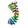
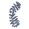
| Structure viewer | Molecule: MolmilJmol/JSmol |
|---|
- Downloads & links
-Download
| PDBx/mmCIF format | 1q1t.cif.gz | 106 KB | Display | PDBx/mmCIF format |
|---|---|---|---|---|
| PDB format | pdb1q1t.ent.gz | 79.1 KB | Display | PDB format |
| PDBx/mmJSON format | 1q1t.json.gz | Tree view | PDBx/mmJSON format | |
| Others |  Other downloads Other downloads |
-Validation report
| Arichive directory | https://data.pdbj.org/pub/pdb/validation_reports/q1/1q1tftp://data.pdbj.org/pub/pdb/validation_reports/q1/1q1t | HTTPS FTP |
|---|
-Related structure data
| Related structure data | 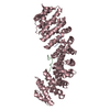 1q1sC 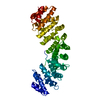 1ialS S: Starting model for refinement C: citing same article ( |
|---|---|
| Similar structure data |


-Links
 PDBj
PDBj


- Assembly
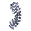
| Deposited unit | 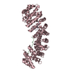
| ||||||||
|---|---|---|---|---|---|---|---|---|---|
| 1 |
| ||||||||
| Unit cell |
|
-Components
| #1: Protein/peptide | Mass: 2685.940 Da / Num. of mol.: 2 / Fragment: CN peptide / Mutation: S120A, S123A, T124A / Source method: obtained synthetically Details: THE PEPTIDE WAS CHEMICALLY SYNTHESIZED. THE SEQUENCE OF THIS PEPTIDE IS NATURALLY FOUND IN SIMIAN VIRUS 40 (SV40) LARGE TUMOR-ANTIGEN References: UniProt: P03070 #2: Protein | | Mass: 50461.285 Da / Num. of mol.: 1 / Fragment: NLS binding domain (70-529) Source method: isolated from a genetically manipulated source Source: (gene. exp.)  #3: Water | ChemComp-HOH / |  Mass: 18.015 Da / Num. of mol.: 302 / Source method: isolated from a natural source / Formula: H2O Mass: 18.015 Da / Num. of mol.: 302 / Source method: isolated from a natural source / Formula: H2O |
|---|
-Experimental details
-Experiment
| Experiment | Method: X-RAY DIFFRACTION / Number of used crystals: 1 |
|---|
- Sample preparation
| Crystal | Density Matthews: 3.17 Å3/Da / Density % sol: 61.24 % | ||||||||||||||||||||||||||||||
|---|---|---|---|---|---|---|---|---|---|---|---|---|---|---|---|---|---|---|---|---|---|---|---|---|---|---|---|---|---|---|---|
| Crystal grow | Temperature: 293 K / Method: vapor diffusion, hanging drop / pH: 6.25 Details: Sodium Citrate, DTT, pH 6.25, VAPOR DIFFUSION, HANGING DROP, temperature 293K | ||||||||||||||||||||||||||||||
| Crystal grow | *PLUS Method: vapor diffusion / PH range low: 6.5 / PH range high: 6 | ||||||||||||||||||||||||||||||
| Components of the solutions | *PLUS
|
-Data collection
| Diffraction | Mean temperature: 100 K |
|---|---|
| Diffraction source | Source: SYNCHROTRON / Site: SSRL  / Beamline: BL7-1 / Wavelength: 0.97 Å / Beamline: BL7-1 / Wavelength: 0.97 Å |
| Detector | Type: MARRESEARCH / Detector: IMAGE PLATE / Date: Apr 13, 2000 |
| Radiation | Protocol: SINGLE WAVELENGTH / Monochromatic (M) / Laue (L): M / Scattering type: x-ray |
| Radiation wavelength | Wavelength: 0.97 Å / Relative weight: 1 |
| Reflection | Resolution: 2.5→99 Å / Num. all: 25902 / Num. obs: 25902 / % possible obs: 97.2 % / Observed criterion σ(I): 0 / Redundancy: 17.1 % / Biso Wilson estimate: 34.3 Å2 / Rmerge(I) obs: 0.078 / Net I/σ(I): 19.8 |
| Reflection shell | Resolution: 2.5→2.59 Å / Rmerge(I) obs: 0.255 / Mean I/σ(I) obs: 3.9 / % possible all: 88.6 |
| Reflection | *PLUS Num. measured all: 443936 |
| Reflection shell | *PLUS % possible obs: 88.6 % |
- Processing
| Software |
| ||||||||||||||||||||||||||||||||||||||||||||||||||||||||||||||||||||||||||||||||
|---|---|---|---|---|---|---|---|---|---|---|---|---|---|---|---|---|---|---|---|---|---|---|---|---|---|---|---|---|---|---|---|---|---|---|---|---|---|---|---|---|---|---|---|---|---|---|---|---|---|---|---|---|---|---|---|---|---|---|---|---|---|---|---|---|---|---|---|---|---|---|---|---|---|---|---|---|---|---|---|---|---|
| Refinement | Method to determine structure: FOURIER SYNTHESIS Starting model: 1IAL Resolution: 2.5→29.62 Å / Rfactor Rfree error: 0.006 / Isotropic thermal model: RESTRAINED / Cross valid method: THROUGHOUT / σ(F): 0 / Stereochemistry target values: Engh & Huber
| ||||||||||||||||||||||||||||||||||||||||||||||||||||||||||||||||||||||||||||||||
| Solvent computation | Solvent model: FLAT MODEL / Bsol: 42.7542 Å2 / ksol: 0.358481 e/Å3 | ||||||||||||||||||||||||||||||||||||||||||||||||||||||||||||||||||||||||||||||||
| Displacement parameters | Biso mean: 39 Å2
| ||||||||||||||||||||||||||||||||||||||||||||||||||||||||||||||||||||||||||||||||
| Refine analyze | Luzzati coordinate error obs: 0.28 Å / Luzzati d res low obs: 5 Å / Luzzati sigma a obs: 0.24 Å | ||||||||||||||||||||||||||||||||||||||||||||||||||||||||||||||||||||||||||||||||
| Refinement step | Cycle: LAST / Resolution: 2.5→29.62 Å
| ||||||||||||||||||||||||||||||||||||||||||||||||||||||||||||||||||||||||||||||||
| Refine LS restraints |
| ||||||||||||||||||||||||||||||||||||||||||||||||||||||||||||||||||||||||||||||||
| LS refinement shell | Resolution: 2.5→2.66 Å / Rfactor Rfree error: 0.017 / Total num. of bins used: 6
| ||||||||||||||||||||||||||||||||||||||||||||||||||||||||||||||||||||||||||||||||
| Xplor file |
| ||||||||||||||||||||||||||||||||||||||||||||||||||||||||||||||||||||||||||||||||
| Refinement | *PLUS Highest resolution: 2.5 Å / Lowest resolution: 30 Å / % reflection Rfree: 10 % | ||||||||||||||||||||||||||||||||||||||||||||||||||||||||||||||||||||||||||||||||
| Solvent computation | *PLUS | ||||||||||||||||||||||||||||||||||||||||||||||||||||||||||||||||||||||||||||||||
| Displacement parameters | *PLUS | ||||||||||||||||||||||||||||||||||||||||||||||||||||||||||||||||||||||||||||||||
| Refine LS restraints | *PLUS
|
