 Movie
Movie Controller


+ Open data

- Basic information
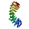
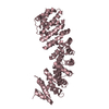
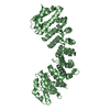
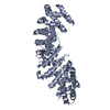
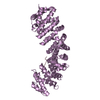
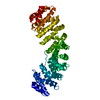
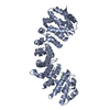
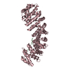
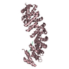
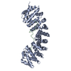
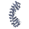
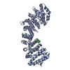
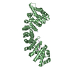
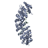
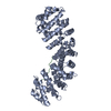
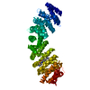
| Entry | Database: PDB / ID: 6d7n | |||||||||
|---|---|---|---|---|---|---|---|---|---|---|
| Title | Crystal structure of the W357R/W399R Importin alpha mutant | |||||||||
 Components Components | Peroxidase,Importin subunit alpha-1 | |||||||||
 Keywords Keywords | TRANSPORT PROTEIN / IMPORTIN / NLS / BIPARTITE / PROTEIN BINDING | |||||||||
| Function / homology |  Function and homology information Function and homology informationSensing of DNA Double Strand Breaks / regulation of transcription by glucose / lactoperoxidase activity / peroxidase / entry of viral genome into host nucleus through nuclear pore complex via importin / positive regulation of viral life cycle / NLS-dependent protein nuclear import complex / postsynapse to nucleus signaling pathway / nuclear import signal receptor activity / nuclear localization sequence binding ...Sensing of DNA Double Strand Breaks / regulation of transcription by glucose / lactoperoxidase activity / peroxidase / entry of viral genome into host nucleus through nuclear pore complex via importin / positive regulation of viral life cycle / NLS-dependent protein nuclear import complex / postsynapse to nucleus signaling pathway / nuclear import signal receptor activity / nuclear localization sequence binding / NLS-bearing protein import into nucleus / non-canonical NF-kappaB signal transduction / positive regulation of type I interferon production / hydrogen peroxide catabolic process / protein import into nucleus / histone deacetylase binding / cytoplasmic stress granule / host cell / response to oxidative stress / nuclear membrane / DNA-binding transcription factor binding / postsynaptic density / heme binding / positive regulation of DNA-templated transcription / glutamatergic synapse / extracellular region / nucleoplasm / metal ion binding / nucleus / cytosol / cytoplasm Similarity search - Function | |||||||||
| Biological species |  Panicum virgatum (switchgrass) Panicum virgatum (switchgrass) | |||||||||
| Method |  X-RAY DIFFRACTION / MOLECULAR REPLACEMENT / Resolution: 2.3 Å X-RAY DIFFRACTION / MOLECULAR REPLACEMENT / Resolution: 2.3 Å | |||||||||
 Authors Authors | Pedersen, L.C. / London, R.E. / Gabel, S.A. | |||||||||
| Funding support |  United States, 2items United States, 2items
| |||||||||
 Citation Citation | Journal: Traffic / Year: 2018 Title: Variations in nuclear localization strategies among pol X family enzymes. Authors: Kirby, T.W. / Pedersen, L.C. / Gabel, S.A. / Gassman, N.R. / London, R.E. | |||||||||
| History |
|
- Structure visualization
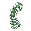
| Structure viewer | Molecule: MolmilJmol/JSmol |
|---|
- Downloads & links
-Download
| PDBx/mmCIF format | 6d7n.cif.gz | 170.1 KB | Display | PDBx/mmCIF format |
|---|---|---|---|---|
| PDB format | pdb6d7n.ent.gz | 132.6 KB | Display | PDB format |
| PDBx/mmJSON format | 6d7n.json.gz | Tree view | PDBx/mmJSON format | |
| Others |  Other downloads Other downloads |
-Validation report
| Arichive directory | https://data.pdbj.org/pub/pdb/validation_reports/d7/6d7nftp://data.pdbj.org/pub/pdb/validation_reports/d7/6d7n | HTTPS FTP |
|---|
-Related structure data
| Related structure data |  5w4eC  5w4fC  6d7mC  5e6qS S: Starting model for refinement C: citing same article ( |
|---|---|
| Similar structure data |

-Links
 PDBj
PDBj


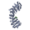
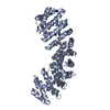
- Assembly
| Deposited unit | 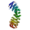
| ||||||||
|---|---|---|---|---|---|---|---|---|---|
| 1 |
| ||||||||
| Unit cell |
|
-Components
| #1: Protein | Mass: 55272.535 Da / Num. of mol.: 1 / Mutation: W357R, W399R Source method: isolated from a genetically manipulated source Source: (gene. exp.) Panicum virgatum (switchgrass), (gene. exp.) Gene: PviPRX9, Kpna2, Rch1 / Production host:  References: UniProt: A0A1S4NYF8, UniProt: P52293, peroxidase | ||
|---|---|---|---|
| #2: Chemical | ChemComp-EDO /   Mass: 62.068 Da / Num. of mol.: 5 / Source method: obtained synthetically / Formula: C2H6O2 Mass: 62.068 Da / Num. of mol.: 5 / Source method: obtained synthetically / Formula: C2H6O2#3: Water | ChemComp-HOH / |  Mass: 18.015 Da / Num. of mol.: 208 / Source method: isolated from a natural source / Formula: H2O Mass: 18.015 Da / Num. of mol.: 208 / Source method: isolated from a natural source / Formula: H2O |
-Experimental details
-Experiment
| Experiment | Method: X-RAY DIFFRACTION / Number of used crystals: 1 |
|---|
- Sample preparation
| Crystal | Density Matthews: 3.24 Å3/Da / Density % sol: 62.05 % |
|---|---|
| Crystal grow | Temperature: 293 K / Method: vapor diffusion, sitting drop / pH: 7 / Details: 1.0M triammonium citrate, 0.1M Bis-Tris-Propane |
-Data collection
| Diffraction | Mean temperature: 100 K |
|---|---|
| Diffraction source | Source: ROTATING ANODE / Type: RIGAKU MICROMAX-007 HF / Wavelength: 1.514 Å |
| Detector | Type: DECTRIS PILATUS 200K / Detector: PIXEL / Date: Mar 28, 2018 / Details: VariMaxHF |
| Radiation | Protocol: SINGLE WAVELENGTH / Monochromatic (M) / Laue (L): M / Scattering type: x-ray |
| Radiation wavelength | Wavelength: 1.514 Å / Relative weight: 1 |
| Reflection | Resolution: 2.3→50 Å / Num. obs: 32563 / % possible obs: 99.9 % / Redundancy: 6.4 % / Rmerge(I) obs: 0.15 / Rpim(I) all: 0.064 / Rrim(I) all: 0.164 / Net I/σ(I): 5.7 |
| Reflection shell | Resolution: 2.3→2.34 Å / Redundancy: 6.1 % / Mean I/σ(I) obs: 2.2 / Num. unique obs: 1571 / CC1/2: 0.749 / Rpim(I) all: 0.395 / Rrim(I) all: 0.979 / Rsym value: 0.89 / % possible all: 100 |
- Processing
| Software |
| |||||||||||||||||||||||||||||||||||||||||||||||||||||||||||||||||||||||||||||||||||||||||||
|---|---|---|---|---|---|---|---|---|---|---|---|---|---|---|---|---|---|---|---|---|---|---|---|---|---|---|---|---|---|---|---|---|---|---|---|---|---|---|---|---|---|---|---|---|---|---|---|---|---|---|---|---|---|---|---|---|---|---|---|---|---|---|---|---|---|---|---|---|---|---|---|---|---|---|---|---|---|---|---|---|---|---|---|---|---|---|---|---|---|---|---|---|
| Refinement | Method to determine structure: MOLECULAR REPLACEMENT Starting model: 5E6Q Resolution: 2.3→29.287 Å / SU ML: 0.25 / Cross valid method: THROUGHOUT / σ(F): 1.43 / Phase error: 22.77
| |||||||||||||||||||||||||||||||||||||||||||||||||||||||||||||||||||||||||||||||||||||||||||
| Solvent computation | Shrinkage radii: 0.9 Å / VDW probe radii: 1.11 Å | |||||||||||||||||||||||||||||||||||||||||||||||||||||||||||||||||||||||||||||||||||||||||||
| Refinement step | Cycle: LAST / Resolution: 2.3→29.287 Å
| |||||||||||||||||||||||||||||||||||||||||||||||||||||||||||||||||||||||||||||||||||||||||||
| Refine LS restraints |
| |||||||||||||||||||||||||||||||||||||||||||||||||||||||||||||||||||||||||||||||||||||||||||
| LS refinement shell |
|
