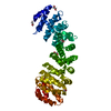
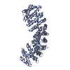
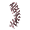
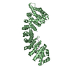
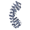
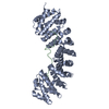
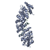
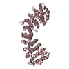
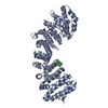
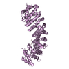
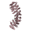
- PDB-6d7m: Crystal structure of the W184R/W231R Importin alpha mutant -
+
データを開く
IDまたはキーワード:
読み込み中...
-
基本情報
登録情報
データベース: PDB / ID: 6d7m
タイトル
Crystal structure of the W184R/W231R Importin alpha mutant
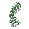
要素
Peroxidase,Importin subunit alpha-1
キーワード
TRANSPORT PROTEIN / IMPORTIN / NLS / BIPARTITE / PROTEIN BINDING
機能・相同性
機能・相同性情報
Sensing of DNA Double Strand Breaks / regulation of transcription by glucose / entry of viral genome into host nucleus through nuclear pore complex via importin / positive regulation of viral life cycle / NLS-dependent protein nuclear import complex / postsynapse to nucleus signaling pathway / nuclear import signal receptor activity / nuclear localization sequence binding / NLS-bearing protein import into nucleus / non-canonical NF-kappaB signal transduction ...Sensing of DNA Double Strand Breaks / regulation of transcription by glucose / entry of viral genome into host nucleus through nuclear pore complex via importin / positive regulation of viral life cycle / NLS-dependent protein nuclear import complex / postsynapse to nucleus signaling pathway / nuclear import signal receptor activity / nuclear localization sequence binding / NLS-bearing protein import into nucleus / non-canonical NF-kappaB signal transduction / positive regulation of type I interferon production / histone deacetylase binding / cytoplasmic stress granule / protein import into nucleus / host cell / nuclear membrane / DNA-binding transcription factor binding / postsynaptic density / positive regulation of DNA-templated transcription / glutamatergic synapse / nucleoplasm / nucleus / cytoplasm / cytosol 類似検索 - 分子機能
 ムービー
ムービー コントローラー


 データを開く
 基本情報
基本情報 要素
要素 キーワード
キーワード 機能・相同性情報
 X線回折 /
  データ登録者
 米国, 2件
  引用
引用 構造の表示
構造の表示 ダウンロードとリンク
 その他のダウンロード


 PDBj
PDBj


 集合体
集合体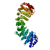


 分子量: 62.068 Da / 分子数: 3 / 由来タイプ: 合成 / 式: C2H6O2
 分子量: 35.453 Da / 分子数: 1 / 由来タイプ: 合成 / 式: Cl
 分子量: 118.088 Da / 分子数: 1 / 由来タイプ: 合成 / 式: C4H6O4
 分子量: 18.015 Da / 分子数: 162 / 由来タイプ: 天然 / 式: H2O
 試料調製
試料調製 解析
解析