- PDB-5ekg: Crystallization and X-ray Diffraction Data Collection of Importin... -
+
Open data
ID or keywords:
Loading...
-
Basic information
Entry
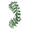
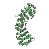
Database: PDB / ID: 5ekg
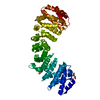
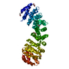
Title
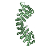
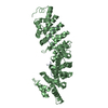
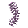
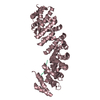
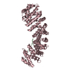
Crystallization and X-ray Diffraction Data Collection of Importin-alpha from Mus musculus Complexed with a XPG NLS Peptide, fragment 2
Components
Importin subunit alpha-1
XPG2 peptide
Keywords
PROTEIN BINDING / Importin alpha / nuclear import pathway / nuclear localization sequence (NLS) / DNA repair proteins / nucleotide excision repair / XPG protein
Function / homology
Function and homology information
nucleotide-excision repair complex / base-excision repair, AP site formation / Sensing of DNA Double Strand Breaks / regulation of transcription by glucose / entry of viral genome into host nucleus through nuclear pore complex via importin / positive regulation of viral life cycle / bubble DNA binding / NLS-dependent protein nuclear import complex / postsynapse to nucleus signaling pathway / hydrolase activity, acting on ester bonds ...nucleotide-excision repair complex / base-excision repair, AP site formation / Sensing of DNA Double Strand Breaks / regulation of transcription by glucose / entry of viral genome into host nucleus through nuclear pore complex via importin / positive regulation of viral life cycle / bubble DNA binding / NLS-dependent protein nuclear import complex / postsynapse to nucleus signaling pathway / hydrolase activity, acting on ester bonds / response to UV-C / nuclear import signal receptor activity / nuclear localization sequence binding / NLS-bearing protein import into nucleus / non-canonical NF-kappaB signal transduction / RNA polymerase II complex binding / response to UV / positive regulation of type I interferon production / transcription-coupled nucleotide-excision repair / nucleotide-excision repair / DNA endonuclease activity / double-strand break repair via homologous recombination / enzyme activator activity / Dual Incision in GG-NER / histone deacetylase binding / cytoplasmic stress granule / Formation of Incision Complex in GG-NER / protein import into nucleus / Dual incision in TC-NER / host cell / single-stranded DNA binding / chromosome / double-stranded DNA binding / endonuclease activity / nuclear membrane / DNA-binding transcription factor binding / Hydrolases; Acting on ester bonds / damaged DNA binding / postsynaptic density / negative regulation of apoptotic process / positive regulation of DNA-templated transcription / protein-containing complex binding / glutamatergic synapse / protein homodimerization activity / protein-containing complex / nucleoplasm / metal ion binding / nucleus / cytoplasm / cytosol Similarity search - Function
In the structure databanks used in Yorodumi, some data are registered as the other names, "COVID-19 virus" and "2019-nCoV". Here are the details of the virus and the list of structure data.
Jan 31, 2019. EMDB accession codes are about to change! (news from PDBe EMDB page)
EMDB accession codes are about to change! (news from PDBe EMDB page)
The allocation of 4 digits for EMDB accession codes will soon come to an end. Whilst these codes will remain in use, new EMDB accession codes will include an additional digit and will expand incrementally as the available range of codes is exhausted. The current 4-digit format prefixed with “EMD-” (i.e. EMD-XXXX) will advance to a 5-digit format (i.e. EMD-XXXXX), and so on. It is currently estimated that the 4-digit codes will be depleted around Spring 2019, at which point the 5-digit format will come into force.
The EM Navigator/Yorodumi systems omit the EMD- prefix.
Related info.:Q: What is EMD? / ID/Accession-code notation in Yorodumi/EM Navigator
Yorodumi is a browser for structure data from EMDB, PDB, SASBDB, etc.
This page is also the successor to EM Navigator detail page, and also detail information page/front-end page for Omokage search.
The word "yorodu" (or yorozu) is an old Japanese word meaning "ten thousand". "mi" (miru) is to see.
Related info.:EMDB / PDB / SASBDB / Comparison of 3 databanks / Yorodumi Search / Aug 31, 2016. New EM Navigator & Yorodumi / Yorodumi Papers / Jmol/JSmol / Function and homology information / Changes in new EM Navigator and Yorodumi
 Movie
Movie Controller

 Yorodumi
 Open data
 Basic information
 Components
 Keywords
 Function and homology information
 X-RAY DIFFRACTION /
  Authors
 Citation
 Structure visualization
 Downloads & links
 Other downloads


 PDBj
PDBj


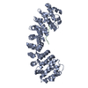
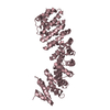
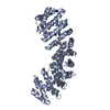
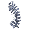
 Assembly
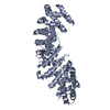

 Sample preparation
 / Beamline: W01B-MX2 / Wavelength: 1.4 Å
 Processing