 Movie
Movie Controller

[English] 日本語
 Yorodumi
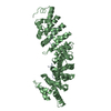
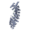
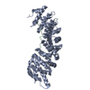
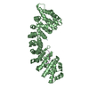
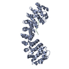
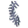
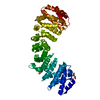
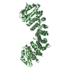
- PDB-5h2x: Crystal structure of the karyopherin Kap60p bound to the SUMO pro... -
+ Open data

- Basic information
| Entry | Database: PDB / ID: 5h2x | ||||||
|---|---|---|---|---|---|---|---|
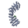
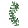
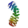
| Title | Crystal structure of the karyopherin Kap60p bound to the SUMO protease Ulp1p (150-172) | ||||||
 Components Components |
| ||||||
 Keywords Keywords | PROTEIN TRANSPORT/HYDROGENASE / nuclear import / PROTEIN TRANSPORT-HYDROGENASE complex | ||||||
| Function / homology |  Function and homology information Function and homology informationUlp1 peptidase / proteasome localization / deSUMOylase activity / protein desumoylation / SUMO is proteolytically processed / import into nucleus / NLS-dependent protein nuclear import complex / NLS-bearing protein import into nucleus / nuclear import signal receptor activity / protein targeting to membrane ...Ulp1 peptidase / proteasome localization / deSUMOylase activity / protein desumoylation / SUMO is proteolytically processed / import into nucleus / NLS-dependent protein nuclear import complex / NLS-bearing protein import into nucleus / nuclear import signal receptor activity / protein targeting to membrane / nuclear localization sequence binding / Major pathway of rRNA processing in the nucleolus and cytosol / nuclear pore / cysteine-type peptidase activity / protein import into nucleus / G2/M transition of mitotic cell cycle / disordered domain specific binding / nuclear envelope / nucleolus / protein-containing complex binding / perinuclear region of cytoplasm / protein-containing complex / proteolysis / nucleus / cytosol / cytoplasm Similarity search - Function | ||||||
| Biological species |  | ||||||
| Method |  X-RAY DIFFRACTION / SYNCHROTRON / MOLECULAR REPLACEMENT / Resolution: 2.2 Å X-RAY DIFFRACTION / SYNCHROTRON / MOLECULAR REPLACEMENT / Resolution: 2.2 Å | ||||||
 Authors Authors | Hirano, H. / Matsuura, Y. | ||||||
| Funding support |  Japan, 1items Japan, 1items
| ||||||
 Citation Citation | Journal: J. Mol. Biol. / Year: 2017 Title: Structures of the Karyopherins Kap121p and Kap60p Bound to the Nuclear Pore-Targeting Domain of the SUMO Protease Ulp1p Authors: Hirano, H. / Kobayashi, J. / Matsuura, Y. | ||||||
| History |
|
- Structure visualization
| Structure viewer | Molecule: MolmilJmol/JSmol |
|---|
- Downloads & links
-Download
| PDBx/mmCIF format | 5h2x.cif.gz | 103.6 KB | Display | PDBx/mmCIF format |
|---|---|---|---|---|
| PDB format | pdb5h2x.ent.gz | 76.6 KB | Display | PDB format |
| PDBx/mmJSON format | 5h2x.json.gz | Tree view | PDBx/mmJSON format | |
| Others |  Other downloads Other downloads |
-Validation report
| Arichive directory | https://data.pdbj.org/pub/pdb/validation_reports/h2/5h2xftp://data.pdbj.org/pub/pdb/validation_reports/h2/5h2x | HTTPS FTP |
|---|
-Related structure data
| Related structure data |  5h2vC  5h2wSC S: Starting model for refinement C: citing same article ( |
|---|---|
| Similar structure data |


-Links
 PDBj
PDBj


- Assembly
| Deposited unit | 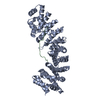
| ||||||||
|---|---|---|---|---|---|---|---|---|---|
| 1 |
| ||||||||
| Unit cell |
|
-Components
| #1: Protein | Mass: 46847.535 Da / Num. of mol.: 1 / Fragment: UNP residues 88-510 Source method: isolated from a genetically manipulated source Source: (gene. exp.) Strain: ATCC 204508 / S288c / Gene: SRP1, KAP60, YNL189W, N1606 / Production host:  |
|---|---|
| #2: Protein/peptide | Mass: 2809.191 Da / Num. of mol.: 1 / Fragment: UNP residues 150-172 Source method: isolated from a genetically manipulated source Source: (gene. exp.) Strain: ATCC 204508 / S288c / Gene: ULP1, YPL020C, LPB11C / Production host: |
| #3: Water | ChemComp-HOH /  Mass: 18.015 Da / Num. of mol.: 220 / Source method: isolated from a natural source / Formula: H2O Mass: 18.015 Da / Num. of mol.: 220 / Source method: isolated from a natural source / Formula: H2O |
-Experimental details
-Experiment
| Experiment | Method: X-RAY DIFFRACTION / Number of used crystals: 1 |
|---|
- Sample preparation
| Crystal | Density Matthews: 2.44 Å3/Da / Density % sol: 49.57 % Description: THE ENTRY CONTAINS FRIEDEL PAIRS IN F_PLUS/MINUS COLUMNS. |
|---|---|
| Crystal grow | Temperature: 293 K / Method: vapor diffusion, hanging drop / pH: 6.5 / Details: 0.1M sodium phosphate, 12% PEG8000 |
-Data collection
| Diffraction | Mean temperature: 100 K | ||||||||||||||||||
|---|---|---|---|---|---|---|---|---|---|---|---|---|---|---|---|---|---|---|---|
| Diffraction source | Source: SYNCHROTRON / Site: Photon Factory / Beamline: BL-17A / Wavelength: 0.98 Å | ||||||||||||||||||
| Detector | Type: DECTRIS PILATUS3 S 6M / Detector: PIXEL / Date: Jun 14, 2015 | ||||||||||||||||||
| Radiation | Protocol: SINGLE WAVELENGTH / Monochromatic (M) / Laue (L): M / Scattering type: x-ray | ||||||||||||||||||
| Radiation wavelength | Wavelength: 0.98 Å / Relative weight: 1 | ||||||||||||||||||
| Reflection | Resolution: 2.2→49.27 Å / Num. obs: 25386 / % possible obs: 100 % / Redundancy: 5.9 % / Biso Wilson estimate: 25.26 Å2 / CC1/2: 0.995 / Rmerge(I) obs: 0.146 / Rpim(I) all: 0.065 / Rrim(I) all: 0.16 / Net I/σ(I): 9.9 / Num. measured all: 149220 / Scaling rejects: 69 | ||||||||||||||||||
| Reflection shell |
|
- Processing
| Software |
| ||||||||||||||||||||||||||||||||||||||||||||||||||||||||||||
|---|---|---|---|---|---|---|---|---|---|---|---|---|---|---|---|---|---|---|---|---|---|---|---|---|---|---|---|---|---|---|---|---|---|---|---|---|---|---|---|---|---|---|---|---|---|---|---|---|---|---|---|---|---|---|---|---|---|---|---|---|---|
| Refinement | Method to determine structure: MOLECULAR REPLACEMENT Starting model: 5H2W Resolution: 2.2→49.268 Å / SU ML: 0.2 / Cross valid method: FREE R-VALUE / σ(F): 1.34 / Phase error: 23.15 Details: SF FILE CONTAINS FRIEDEL PAIRS UNDER I/F_MINUS AND I/F_PLUS COLUMNS.
| ||||||||||||||||||||||||||||||||||||||||||||||||||||||||||||
| Solvent computation | Shrinkage radii: 0.9 Å / VDW probe radii: 1.11 Å | ||||||||||||||||||||||||||||||||||||||||||||||||||||||||||||
| Displacement parameters | Biso max: 78.79 Å2 / Biso mean: 32.3559 Å2 / Biso min: 13.42 Å2 | ||||||||||||||||||||||||||||||||||||||||||||||||||||||||||||
| Refinement step | Cycle: final / Resolution: 2.2→49.268 Å
| ||||||||||||||||||||||||||||||||||||||||||||||||||||||||||||
| Refine LS restraints |
| ||||||||||||||||||||||||||||||||||||||||||||||||||||||||||||
| LS refinement shell | Refine-ID: X-RAY DIFFRACTION / Total num. of bins used: 9 / % reflection obs: 100 %
|
