 Movie
Movie Controller


+ Open data

- Basic information
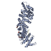
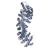
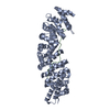
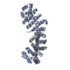
| Entry | Database: PDB / ID: 1bk6 | ||||||
|---|---|---|---|---|---|---|---|
| Title | KARYOPHERIN ALPHA (YEAST) + SV40 T ANTIGEN NLS | ||||||
 Components Components |
| ||||||
 Keywords Keywords | COMPLEX (PROTEIN TRANSPORT/PEPTIDE) / PROTEIN TRANSPORT / NLS NUCLEAR IMPORT / ARMADILLO REPEAT CONTAINING PROTEIN / COMPLEX (PROTEIN TRANSPORT-PEPTIDE) / COMPLEX (PROTEIN TRANSPORT-PEPTIDE) complex | ||||||
| Function / homology |  Function and homology information Function and homology informationproteasome localization / import into nucleus / NLS-dependent protein nuclear import complex / nuclear import signal receptor activity / protein targeting to membrane / nuclear localization sequence binding / NLS-bearing protein import into nucleus / disordered domain specific binding / protein import into nucleus / nuclear envelope ...proteasome localization / import into nucleus / NLS-dependent protein nuclear import complex / nuclear import signal receptor activity / protein targeting to membrane / nuclear localization sequence binding / NLS-bearing protein import into nucleus / disordered domain specific binding / protein import into nucleus / nuclear envelope / protein-containing complex binding / perinuclear region of cytoplasm / protein-containing complex / nucleoplasm / nucleus / cytoplasm / cytosol Similarity search - Function | ||||||
| Biological species |  | ||||||
| Method |  X-RAY DIFFRACTION / SYNCHROTRON / DIFFERENCE FOURIER / Resolution: 2.8 Å X-RAY DIFFRACTION / SYNCHROTRON / DIFFERENCE FOURIER / Resolution: 2.8 Å | ||||||
 Authors Authors | Conti, E. / Uy, M. / Leighton, L. / Blobel, G. / Kuriyan, J. | ||||||
 Citation Citation | Journal: Cell(Cambridge,Mass.) / Year: 1998 Title: Crystallographic analysis of the recognition of a nuclear localization signal by the nuclear import factor karyopherin alpha. Authors: Conti, E. / Uy, M. / Leighton, L. / Blobel, G. / Kuriyan, J. | ||||||
| History |
|
- Structure visualization
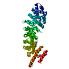
| Structure viewer | Molecule: MolmilJmol/JSmol |
|---|
- Downloads & links
-Download
| PDBx/mmCIF format | 1bk6.cif.gz | 166.4 KB | Display | PDBx/mmCIF format |
|---|---|---|---|---|
| PDB format | pdb1bk6.ent.gz | 132 KB | Display | PDB format |
| PDBx/mmJSON format | 1bk6.json.gz | Tree view | PDBx/mmJSON format | |
| Others |  Other downloads Other downloads |
-Validation report
| Arichive directory | https://data.pdbj.org/pub/pdb/validation_reports/bk/1bk6ftp://data.pdbj.org/pub/pdb/validation_reports/bk/1bk6 | HTTPS FTP |
|---|
-Related structure data


-Links
 PDBj
PDBj


- Assembly
| Deposited unit | 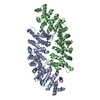
| ||||||||
|---|---|---|---|---|---|---|---|---|---|
| 1 | 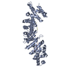
| ||||||||
| 2 | 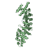
| ||||||||
| Unit cell |
|
-Components
| #1: Protein | Mass: 46718.426 Da / Num. of mol.: 2 / Fragment: ARMADILLO DOMAIN Source method: isolated from a genetically manipulated source Source: (gene. exp.) Strain: W303 / Cell line: BL21 / Cellular location: CYTOPLASMIC/NUCLEAR / Gene: SRP1 / Plasmid: PPROEX-HTB / Species (production host): Escherichia coli / Production host:  #2: Protein/peptide | Mass: 791.059 Da / Num. of mol.: 2 / Fragment: NLS (NUCLEAR LOCALIZATION SIGNAL) Source method: isolated from a genetically manipulated source Details: NLS PEPTIDE AT THE LARGER (FUNCTIONAL) BINDING SITE #3: Protein/peptide | Mass: 489.608 Da / Num. of mol.: 2 / Fragment: NLS (NUCLEAR LOCALIZATION SIGNAL) Source method: isolated from a genetically manipulated source Details: NLS PEPTIDE AT THE SMALLER BINDING SITE #4: Water | ChemComp-HOH / |  Mass: 18.015 Da / Num. of mol.: 12 / Source method: isolated from a natural source / Formula: H2O Mass: 18.015 Da / Num. of mol.: 12 / Source method: isolated from a natural source / Formula: H2O |
|---|
-Experimental details
-Experiment
| Experiment | Method: X-RAY DIFFRACTION / Number of used crystals: 1 |
|---|
- Sample preparation
| Crystal | Density Matthews: 2.6 Å3/Da / Density % sol: 50 % | ||||||||||||||||||||||||||||||
|---|---|---|---|---|---|---|---|---|---|---|---|---|---|---|---|---|---|---|---|---|---|---|---|---|---|---|---|---|---|---|---|
| Crystal grow | pH: 7 / Details: pH 7.0 | ||||||||||||||||||||||||||||||
| Crystal grow | *PLUS Temperature: 4 ℃ / Method: vapor diffusion, hanging drop | ||||||||||||||||||||||||||||||
| Components of the solutions | *PLUS
|
-Data collection
| Diffraction | Mean temperature: 100 K |
|---|---|
| Diffraction source | Source: SYNCHROTRON / Site: CHESS  / Beamline: A1 / Wavelength: 0.9 / Beamline: A1 / Wavelength: 0.9 |
| Detector | Type: BRANDEIS / Detector: CCD / Date: Dec 1, 1997 |
| Radiation | Monochromatic (M) / Laue (L): M / Scattering type: x-ray |
| Radiation wavelength | Wavelength: 0.9 Å / Relative weight: 1 |
| Reflection | Resolution: 2.8→29.1 Å / Num. obs: 22717 / % possible obs: 86.9 % / Redundancy: 3.5 % / Rsym value: 0.064 / Net I/σ(I): 17.4 |
| Reflection shell | Resolution: 2.8→2.9 Å / Redundancy: 2.4 % / Mean I/σ(I) obs: 9.9 / Rsym value: 0.188 / % possible all: 82.2 |
| Reflection | *PLUS Num. measured all: 270860 / Rmerge(I) obs: 0.064 |
| Reflection shell | *PLUS % possible obs: 82.2 % / Rmerge(I) obs: 0.188 |
- Processing
| Software |
| ||||||||||||||||||||||||||||||||||||||||||||||||||||||||||||
|---|---|---|---|---|---|---|---|---|---|---|---|---|---|---|---|---|---|---|---|---|---|---|---|---|---|---|---|---|---|---|---|---|---|---|---|---|---|---|---|---|---|---|---|---|---|---|---|---|---|---|---|---|---|---|---|---|---|---|---|---|---|
| Refinement | Method to determine structure: DIFFERENCE FOURIER Starting model: UNLIGANDED KARYOPHERIN ALPHA Resolution: 2.8→29.1 Å / Isotropic thermal model: RESTRAINED / Cross valid method: THROUGHOUT / σ(F): 0
| ||||||||||||||||||||||||||||||||||||||||||||||||||||||||||||
| Solvent computation | Solvent model: FLAT MODEL | ||||||||||||||||||||||||||||||||||||||||||||||||||||||||||||
| Displacement parameters |
| ||||||||||||||||||||||||||||||||||||||||||||||||||||||||||||
| Refinement step | Cycle: LAST / Resolution: 2.8→29.1 Å
| ||||||||||||||||||||||||||||||||||||||||||||||||||||||||||||
| Refine LS restraints |
| ||||||||||||||||||||||||||||||||||||||||||||||||||||||||||||
| LS refinement shell | Resolution: 2.8→2.85 Å / Total num. of bins used: 18
| ||||||||||||||||||||||||||||||||||||||||||||||||||||||||||||
| Xplor file |
| ||||||||||||||||||||||||||||||||||||||||||||||||||||||||||||
| Software | *PLUS Name: CNS / Version: 0.2 / Classification: refinement | ||||||||||||||||||||||||||||||||||||||||||||||||||||||||||||
| Refinement | *PLUS | ||||||||||||||||||||||||||||||||||||||||||||||||||||||||||||
| Solvent computation | *PLUS | ||||||||||||||||||||||||||||||||||||||||||||||||||||||||||||
| Displacement parameters | *PLUS | ||||||||||||||||||||||||||||||||||||||||||||||||||||||||||||
| Refine LS restraints | *PLUS
| ||||||||||||||||||||||||||||||||||||||||||||||||||||||||||||
| LS refinement shell | *PLUS Rfactor obs: 0.296 |
