 Movie
Movie Controller

[English] 日本語
 Yorodumi
- PDB-1un0: Crystal Structure of Yeast Karyopherin (Importin) alpha in comple... -
+ Open data

- Basic information
| Entry | Database: PDB / ID: 1un0 | ||||||
|---|---|---|---|---|---|---|---|
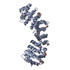
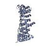
| Title | Crystal Structure of Yeast Karyopherin (Importin) alpha in complex with a Nup2p N-terminal fragment | ||||||
 Components Components |
| ||||||
 Keywords Keywords | NUCLEAR IMPORT / ARMADILLO REPEAT / NUCLEOPORIN / NLS RELEASE / KARYOPHERIN RECYCLING | ||||||
| Function / homology |  Function and homology information Function and homology informationproteasome localization / mRNA export from nucleus in response to heat stress / transcription-dependent tethering of RNA polymerase II gene DNA at nuclear periphery / protein localization to nuclear inner membrane / import into nucleus / nuclear pore cytoplasmic filaments / post-transcriptional tethering of RNA polymerase II gene DNA at nuclear periphery / nuclear pore nuclear basket / NLS-dependent protein nuclear import complex / importin-alpha family protein binding ...proteasome localization / mRNA export from nucleus in response to heat stress / transcription-dependent tethering of RNA polymerase II gene DNA at nuclear periphery / protein localization to nuclear inner membrane / import into nucleus / nuclear pore cytoplasmic filaments / post-transcriptional tethering of RNA polymerase II gene DNA at nuclear periphery / nuclear pore nuclear basket / NLS-dependent protein nuclear import complex / importin-alpha family protein binding / structural constituent of nuclear pore / silent mating-type cassette heterochromatin formation / nuclear import signal receptor activity / protein targeting to membrane / nucleocytoplasmic transport / nuclear localization sequence binding / NLS-bearing protein import into nucleus / poly(A)+ mRNA export from nucleus / subtelomeric heterochromatin formation / nuclear pore / protein export from nucleus / protein import into nucleus / small GTPase binding / disordered domain specific binding / nuclear envelope / nuclear membrane / chromosome, telomeric region / protein-containing complex binding / perinuclear region of cytoplasm / protein-containing complex / mitochondrion / nucleoplasm / nucleus / cytosol / cytoplasm Similarity search - Function | ||||||
| Biological species |  | ||||||
| Method |  X-RAY DIFFRACTION / SYNCHROTRON / MOLECULAR REPLACEMENT / Resolution: 2.6 Å X-RAY DIFFRACTION / SYNCHROTRON / MOLECULAR REPLACEMENT / Resolution: 2.6 Å | ||||||
 Authors Authors | Matsuura, Y. / Lange, A. / Harreman, M.T. / Corbett, A.H. / Stewart, M. | ||||||
 Citation Citation | Journal: Embo J. / Year: 2003 Title: Structural Basis for Nup2P Function in Cargo Release and Karyopherin Recycling in Nuclear Import Authors: Matsuura, Y. / Lange, A. / Harreman, M.T. / Corbett, A.H. / Stewart, M. | ||||||
| History |
|
- Structure visualization
| Structure viewer | Molecule: MolmilJmol/JSmol |
|---|
- Downloads & links
-Download
| PDBx/mmCIF format | 1un0.cif.gz | 188.8 KB | Display | PDBx/mmCIF format |
|---|---|---|---|---|
| PDB format | pdb1un0.ent.gz | 149.7 KB | Display | PDB format |
| PDBx/mmJSON format | 1un0.json.gz | Tree view | PDBx/mmJSON format | |
| Others |  Other downloads Other downloads |
-Validation report
| Arichive directory | https://data.pdbj.org/pub/pdb/validation_reports/un/1un0ftp://data.pdbj.org/pub/pdb/validation_reports/un/1un0 | HTTPS FTP |
|---|
-Related structure data
| Related structure data | 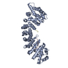 1ee4S S: Starting model for refinement |
|---|---|
| Similar structure data |


-Links
 PDBj
PDBj


- Assembly
| Deposited unit | 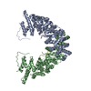
| ||||||||
|---|---|---|---|---|---|---|---|---|---|
| 1 | 
| ||||||||
| 2 | 
| ||||||||
| Unit cell |
|
-Components
| #1: Protein | Mass: 48906.562 Da / Num. of mol.: 2 / Fragment: ARMADILLO REPEAT DOMAIN, RESIDUES 88-530 / Mutation: YES Source method: isolated from a genetically manipulated source Source: (gene. exp.) Plasmid: PET30A / Production host:  #2: Protein | Mass: 5813.649 Da / Num. of mol.: 2 / Fragment: N-TERMINAL FRAGMENT, RESIDUES 1-51 Source method: isolated from a genetically manipulated source Source: (gene. exp.) Plasmid: PET30A / Production host: #3: Water | ChemComp-HOH / |  Mass: 18.015 Da / Num. of mol.: 158 / Source method: isolated from a natural source / Formula: H2O Mass: 18.015 Da / Num. of mol.: 158 / Source method: isolated from a natural source / Formula: H2OCompound details | CHAIN A, B ENGINEERED | |
|---|
-Experimental details
-Experiment
| Experiment | Method: X-RAY DIFFRACTION / Number of used crystals: 1 |
|---|
- Sample preparation
| Crystal | Density Matthews: 2.4 Å3/Da / Density % sol: 49 % | ||||||||||||||||||||||||||||||||||||||||||
|---|---|---|---|---|---|---|---|---|---|---|---|---|---|---|---|---|---|---|---|---|---|---|---|---|---|---|---|---|---|---|---|---|---|---|---|---|---|---|---|---|---|---|---|
| Crystal grow | pH: 6.8 Details: 50 MM HEPES PH 6.8, 0.15 M NACL, 24 % PEG3350, 2 % PEG400 | ||||||||||||||||||||||||||||||||||||||||||
| Crystal grow | *PLUS Temperature: 18 ℃ / pH: 6.8 / Method: vapor diffusion, hanging drop | ||||||||||||||||||||||||||||||||||||||||||
| Components of the solutions | *PLUS
|
-Data collection
| Diffraction | Mean temperature: 100 K |
|---|---|
| Diffraction source | Source: SYNCHROTRON / Site: SRS  / Beamline: PX14.2 / Wavelength: 0.978 / Beamline: PX14.2 / Wavelength: 0.978 |
| Detector | Type: ADSC CCD / Detector: CCD / Date: Apr 21, 2002 |
| Radiation | Protocol: SINGLE WAVELENGTH / Monochromatic (M) / Laue (L): M / Scattering type: x-ray |
| Radiation wavelength | Wavelength: 0.978 Å / Relative weight: 1 |
| Reflection | Resolution: 2.6→20 Å / Num. obs: 29670 / % possible obs: 97.5 % / Redundancy: 2.3 % / Rmerge(I) obs: 0.086 / Net I/σ(I): 10 |
| Reflection shell | Resolution: 2.6→2.74 Å / Redundancy: 2.3 % / Rmerge(I) obs: 0.531 / Mean I/σ(I) obs: 1.8 / % possible all: 98.3 |
| Reflection | *PLUS Highest resolution: 2.6 Å / Lowest resolution: 20 Å / Num. measured all: 76810 / Rmerge(I) obs: 0.086 |
| Reflection shell | *PLUS % possible obs: 98.3 % / Num. unique obs: 4308 / Num. measured obs: 10875 / Rmerge(I) obs: 0.531 / Mean I/σ(I) obs: 1.8 |
- Processing
| Software |
| ||||||||||||||||||||
|---|---|---|---|---|---|---|---|---|---|---|---|---|---|---|---|---|---|---|---|---|---|
| Refinement | Method to determine structure: MOLECULAR REPLACEMENT Starting model: PDB ENTRY 1EE4 Resolution: 2.6→20 Å / SU B: 11.435 / SU ML: 0.242 / Cross valid method: THROUGHOUT / ESU R: 0.686 / ESU R Free: 0.316
| ||||||||||||||||||||
| Displacement parameters | Biso mean: 49.22 Å2
| ||||||||||||||||||||
| Refinement step | Cycle: LAST / Resolution: 2.6→20 Å
| ||||||||||||||||||||
| Refinement | *PLUS Highest resolution: 2.6 Å / Lowest resolution: 20 Å / Rfactor Rfree: 0.257 / Rfactor Rwork: 0.216 | ||||||||||||||||||||
| Solvent computation | *PLUS | ||||||||||||||||||||
| Displacement parameters | *PLUS | ||||||||||||||||||||
| Refine LS restraints | *PLUS
|
