 Movie
Movie Controller


+ Open data

- Basic information
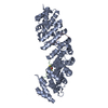
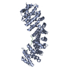
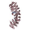
| Entry | Database: PDB / ID: 1ejy | ||||||
|---|---|---|---|---|---|---|---|
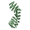
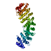
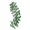
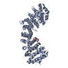
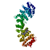
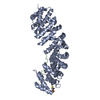
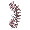
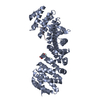
| Title | MOUSE IMPORTIN ALPHA-NUCLEOPLASMIN NLS PEPTIDE COMPLEX | ||||||
 Components Components |
| ||||||
 Keywords Keywords | Hydrolase/Peptide / importin aplpha/karyopherin alpha / nuclear localization sequence (NLS) recognition / nucleoplasmin / PROTEIN BINDING / Hydrolase-Peptide complex | ||||||
| Function / homology |  Function and homology information Function and homology informationsperm DNA decondensation / histone chaperone activity / Sensing of DNA Double Strand Breaks / regulation of transcription by glucose / entry of viral genome into host nucleus through nuclear pore complex via importin / positive regulation of viral life cycle / importin-alpha family protein binding / NLS-dependent protein nuclear import complex / postsynapse to nucleus signaling pathway / nuclear import signal receptor activity ...sperm DNA decondensation / histone chaperone activity / Sensing of DNA Double Strand Breaks / regulation of transcription by glucose / entry of viral genome into host nucleus through nuclear pore complex via importin / positive regulation of viral life cycle / importin-alpha family protein binding / NLS-dependent protein nuclear import complex / postsynapse to nucleus signaling pathway / nuclear import signal receptor activity / nuclear localization sequence binding / NLS-bearing protein import into nucleus / non-canonical NF-kappaB signal transduction / nucleosome binding / positive regulation of type I interferon production / positive regulation of DNA replication / histone deacetylase binding / cytoplasmic stress granule / protein import into nucleus / host cell / histone binding / nuclear membrane / DNA-binding transcription factor binding / postsynaptic density / chromatin remodeling / chromatin binding / positive regulation of DNA-templated transcription / nucleolus / glutamatergic synapse / RNA binding / nucleoplasm / nucleus / cytoplasm / cytosol Similarity search - Function | ||||||
| Biological species |  | ||||||
| Method |  X-RAY DIFFRACTION / Resolution: 2.9 Å X-RAY DIFFRACTION / Resolution: 2.9 Å | ||||||
 Authors Authors | Fontes, M.R. / Teh, T. / Kobe, B. | ||||||
 Citation Citation | Journal: J.Mol.Biol. / Year: 2000 Title: Structural basis of recognition of monopartite and bipartite nuclear localization sequences by mammalian importin-alpha. Authors: Fontes, M.R. / Teh, T. / Kobe, B. #1: Journal: Nat.Struct.Biol. / Year: 1999Title: Autoinhibition by an Internal Nuclear Localization Signal Reveled by the Crystal Structure of Mammalian Importin Alpha Authors: Kobe, B. | ||||||
| History |
|
- Structure visualization
| Structure viewer | Molecule: MolmilJmol/JSmol |
|---|
- Downloads & links
-Download
| PDBx/mmCIF format | 1ejy.cif.gz | 94.3 KB | Display | PDBx/mmCIF format |
|---|---|---|---|---|
| PDB format | pdb1ejy.ent.gz | 71.4 KB | Display | PDB format |
| PDBx/mmJSON format | 1ejy.json.gz | Tree view | PDBx/mmJSON format | |
| Others |  Other downloads Other downloads |
-Validation report
| Arichive directory | https://data.pdbj.org/pub/pdb/validation_reports/ej/1ejyftp://data.pdbj.org/pub/pdb/validation_reports/ej/1ejy | HTTPS FTP |
|---|
-Related structure data


-Links
 PDBj
PDBj


- Assembly
| Deposited unit | 
| ||||||||
|---|---|---|---|---|---|---|---|---|---|
| 1 |
| ||||||||
| Unit cell |
|
-Components
| #1: Protein/peptide | Mass: 1747.178 Da / Num. of mol.: 1 Fragment: NLS (NUCLEAR LOCALIZATION SIGNAL) BIPARTITE PEPTIDE, RESIDUES 155-170 Source method: obtained synthetically Details: The peptide was chemically synthesized. The sequence of this peptide is naturally found in Xenopus laevis (African clawed frog). References: UniProt: P05221 |
|---|---|
| #2: Protein | Mass: 49886.633 Da / Num. of mol.: 1 / Fragment: NLS BINDING DOMAIN, RESIDUES 70-529 Source method: isolated from a genetically manipulated source Source: (gene. exp.)  |
-Experimental details
-Experiment
| Experiment | Method: X-RAY DIFFRACTION / Number of used crystals: 1 |
|---|
- Sample preparation
| Crystal | Density Matthews: 3.43 Å3/Da / Density % sol: 64.1 % | ||||||||||||||||||||
|---|---|---|---|---|---|---|---|---|---|---|---|---|---|---|---|---|---|---|---|---|---|
| Crystal grow | Temperature: 293 K / Method: vapor diffusion, hanging drop / pH: 6.5 Details: sodium citrate, Hepes, DTT, pH 6.5, VAPOR DIFFUSION, HANGING DROP, temperature 293K | ||||||||||||||||||||
| Crystal grow | *PLUS Method: unknown | ||||||||||||||||||||
| Components of the solutions | *PLUS
|
-Data collection
| Diffraction | Mean temperature: 100 K |
|---|---|
| Diffraction source | Source: ROTATING ANODE / Type: RIGAKU RU200 / Wavelength: 1.5418 |
| Detector | Type: MARRESEARCH / Detector: AREA DETECTOR / Date: Sep 9, 1999 |
| Radiation | Protocol: SINGLE WAVELENGTH / Monochromatic (M) / Laue (L): M / Scattering type: x-ray |
| Radiation wavelength | Wavelength: 1.5418 Å / Relative weight: 1 |
| Reflection | Resolution: 2.9→30 Å / Num. all: 121070 / Num. obs: 16370 / % possible obs: 98.9 % / Observed criterion σ(F): 0 / Observed criterion σ(I): -3 / Redundancy: 7.4 % / Biso Wilson estimate: 68.3 Å2 / Rmerge(I) obs: 0.239 / Net I/σ(I): 7.2 |
| Reflection shell | Resolution: 2.9→3 Å / Redundancy: 6.1 % / Rmerge(I) obs: 0.962 / Num. unique all: 1598 / % possible all: 100 |
| Reflection | *PLUS Num. measured all: 121070 |
| Reflection shell | *PLUS % possible obs: 100 % / Num. unique obs: 1598 / Mean I/σ(I) obs: 1.3 |
- Processing
| Software |
| ||||||||||||||||||||||||||||||||||||||||||||||||||||||||||||||||||||||||||||||||
|---|---|---|---|---|---|---|---|---|---|---|---|---|---|---|---|---|---|---|---|---|---|---|---|---|---|---|---|---|---|---|---|---|---|---|---|---|---|---|---|---|---|---|---|---|---|---|---|---|---|---|---|---|---|---|---|---|---|---|---|---|---|---|---|---|---|---|---|---|---|---|---|---|---|---|---|---|---|---|---|---|---|
| Refinement | Resolution: 2.9→30 Å / Rfactor Rfree error: 0.007 / Data cutoff high absF: 1331801.17 / Data cutoff low absF: 0 / Isotropic thermal model: RESTRAINED / Cross valid method: THROUGHOUT / σ(F): 0 / σ(I): 0 / Stereochemistry target values: protein_rep.param
| ||||||||||||||||||||||||||||||||||||||||||||||||||||||||||||||||||||||||||||||||
| Solvent computation | Solvent model: FLAT MODEL / Bsol: 28.39 Å2 / ksol: 0.371 e/Å3 | ||||||||||||||||||||||||||||||||||||||||||||||||||||||||||||||||||||||||||||||||
| Displacement parameters | Biso mean: 37.2 Å2
| ||||||||||||||||||||||||||||||||||||||||||||||||||||||||||||||||||||||||||||||||
| Refine analyze |
| ||||||||||||||||||||||||||||||||||||||||||||||||||||||||||||||||||||||||||||||||
| Refinement step | Cycle: LAST / Resolution: 2.9→30 Å
| ||||||||||||||||||||||||||||||||||||||||||||||||||||||||||||||||||||||||||||||||
| Refine LS restraints |
| ||||||||||||||||||||||||||||||||||||||||||||||||||||||||||||||||||||||||||||||||
| LS refinement shell | Resolution: 2.9→3 Å / Rfactor Rfree error: 0.029 / Total num. of bins used: 10
| ||||||||||||||||||||||||||||||||||||||||||||||||||||||||||||||||||||||||||||||||
| Xplor file | Serial no: 1 / Param file: PROTEIN_REP.PARAM / Topol file: PROTEIN.TOP | ||||||||||||||||||||||||||||||||||||||||||||||||||||||||||||||||||||||||||||||||
| Software | *PLUS Name: CNS / Version: 0.4 / Classification: refinement | ||||||||||||||||||||||||||||||||||||||||||||||||||||||||||||||||||||||||||||||||
| Refinement | *PLUS σ(F): 0 / % reflection Rfree: 9.8 % | ||||||||||||||||||||||||||||||||||||||||||||||||||||||||||||||||||||||||||||||||
| Solvent computation | *PLUS | ||||||||||||||||||||||||||||||||||||||||||||||||||||||||||||||||||||||||||||||||
| Displacement parameters | *PLUS | ||||||||||||||||||||||||||||||||||||||||||||||||||||||||||||||||||||||||||||||||
| Refine LS restraints | *PLUS
| ||||||||||||||||||||||||||||||||||||||||||||||||||||||||||||||||||||||||||||||||
| LS refinement shell | *PLUS Rfactor Rfree: 0.355 / % reflection Rfree: 9.5 % / Rfactor Rwork: 0.345 / Rfactor obs: 0.345 |
