 Movie
Movie Controller

[English] 日本語
 Yorodumi
- PDB-5u5r: Crystal Structure and X-ray Diffraction Data Collection of Import... -
+ Open data

- Basic information
| Entry | Database: PDB / ID: 5u5r | ||||||
|---|---|---|---|---|---|---|---|
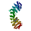
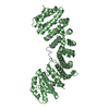
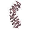
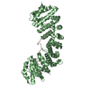
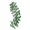
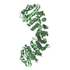
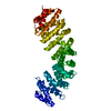
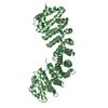
| Title | Crystal Structure and X-ray Diffraction Data Collection of Importin-alpha from Mus musculus Complexed with a PMS2 NLS Peptide | ||||||
 Components Components |
| ||||||
 Keywords Keywords | TRANSPORT PROTEIN / Nuclear transport / Importin-alpha / PMS2 / NLS | ||||||
| Function / homology |  Function and homology information Function and homology informationsingle base insertion or deletion binding / Defective Mismatch Repair Associated With MLH1 / Defective Mismatch Repair Associated With PMS2 / MutLalpha complex / somatic recombination of immunoglobulin gene segments / positive regulation of isotype switching to IgA isotypes / Sensing of DNA Double Strand Breaks / regulation of transcription by glucose / positive regulation of isotype switching to IgG isotypes / entry of viral genome into host nucleus through nuclear pore complex via importin ...single base insertion or deletion binding / Defective Mismatch Repair Associated With MLH1 / Defective Mismatch Repair Associated With PMS2 / MutLalpha complex / somatic recombination of immunoglobulin gene segments / positive regulation of isotype switching to IgA isotypes / Sensing of DNA Double Strand Breaks / regulation of transcription by glucose / positive regulation of isotype switching to IgG isotypes / entry of viral genome into host nucleus through nuclear pore complex via importin / positive regulation of viral life cycle / NLS-dependent protein nuclear import complex / postsynapse to nucleus signaling pathway / Mismatch repair (MMR) directed by MSH2:MSH3 (MutSbeta) / Mismatch repair (MMR) directed by MSH2:MSH6 (MutSalpha) / nuclear import signal receptor activity / nuclear localization sequence binding / NLS-bearing protein import into nucleus / non-canonical NF-kappaB signal transduction / ATP-dependent DNA damage sensor activity / mismatch repair / somatic hypermutation of immunoglobulin genes / positive regulation of type I interferon production / TP53 Regulates Transcription of DNA Repair Genes / histone deacetylase binding / cytoplasmic stress granule / protein import into nucleus / host cell / endonuclease activity / nuclear membrane / DNA-binding transcription factor binding / Hydrolases; Acting on ester bonds / postsynaptic density / response to xenobiotic stimulus / positive regulation of DNA-templated transcription / glutamatergic synapse / ATP hydrolysis activity / DNA binding / nucleoplasm / ATP binding / nucleus / cytoplasm / cytosol Similarity search - Function | ||||||
| Biological species |   Homo sapiens (human) Homo sapiens (human) | ||||||
| Method |  X-RAY DIFFRACTION / SYNCHROTRON / FOURIER SYNTHESIS / Resolution: 2.1 Å X-RAY DIFFRACTION / SYNCHROTRON / FOURIER SYNTHESIS / Resolution: 2.1 Å | ||||||
 Authors Authors | Barros, A.C. / Takeda, A.A. / Dreyer, T.R. / Velazquez-Campoy, A. / Kobe, B. / Fontes, M.R. | ||||||
 Citation Citation | Journal: Biochimie / Year: 2018 Title: DNA mismatch repair proteins MLH1 and PMS2 can be imported to the nucleus by a classical nuclear import pathway. Authors: de Barros, A.C. / Takeda, A.A.S. / Dreyer, T.R. / Velazquez-Campoy, A. / Kobe, B. / Fontes, M.R.M. | ||||||
| History |
|
- Structure visualization
| Structure viewer | Molecule: MolmilJmol/JSmol |
|---|
- Downloads & links
-Download
| PDBx/mmCIF format | 5u5r.cif.gz | 107.8 KB | Display | PDBx/mmCIF format |
|---|---|---|---|---|
| PDB format | pdb5u5r.ent.gz | 78.6 KB | Display | PDB format |
| PDBx/mmJSON format | 5u5r.json.gz | Tree view | PDBx/mmJSON format | |
| Others |  Other downloads Other downloads |
-Validation report
| Arichive directory | https://data.pdbj.org/pub/pdb/validation_reports/u5/5u5rftp://data.pdbj.org/pub/pdb/validation_reports/u5/5u5r | HTTPS FTP |
|---|
-Related structure data
| Related structure data |  5u5pC  1q1sS C: citing same article ( S: Starting model for refinement |
|---|---|
| Similar structure data |


-Links
 PDBj
PDBj


- Assembly
| Deposited unit | 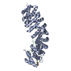
| ||||||||
|---|---|---|---|---|---|---|---|---|---|
| 1 |
| ||||||||
| Unit cell |
|
-Components
| #1: Protein | Mass: 55330.566 Da / Num. of mol.: 1 Source method: isolated from a genetically manipulated source Source: (gene. exp.)  |
|---|---|
| #2: Protein/peptide | Mass: 1381.575 Da / Num. of mol.: 1 / Source method: obtained synthetically / Source: (synth.) Homo sapiens (human)References: UniProt: P54278, Hydrolases; Acting on ester bonds |
| #3: Chemical | ChemComp-DTT /   Mass: 154.251 Da / Num. of mol.: 1 / Source method: obtained synthetically / Formula: C4H10O2S2 Mass: 154.251 Da / Num. of mol.: 1 / Source method: obtained synthetically / Formula: C4H10O2S2 |
| #4: Water | ChemComp-HOH /  Mass: 18.015 Da / Num. of mol.: 332 / Source method: isolated from a natural source / Formula: H2O Mass: 18.015 Da / Num. of mol.: 332 / Source method: isolated from a natural source / Formula: H2O |
-Experimental details
-Experiment
| Experiment | Method: X-RAY DIFFRACTION / Number of used crystals: 1 |
|---|
- Sample preparation
| Crystal | Density Matthews: 2.95 Å3/Da / Density % sol: 58.36 % |
|---|---|
| Crystal grow | Temperature: 291 K / Method: vapor diffusion, hanging drop / Details: 0.600-0.625 M sodium citrate (pH 6) and 10 mM DTT |
-Data collection
| Diffraction | Mean temperature: 100 K |
|---|---|
| Diffraction source | Source: SYNCHROTRON / Site: LNLS  / Beamline: W01B-MX2 / Wavelength: 1 / Beamline: W01B-MX2 / Wavelength: 1 |
| Detector | Type: MARMOSAIC 225 mm CCD / Detector: CCD / Date: Aug 1, 2012 |
| Radiation | Protocol: SINGLE WAVELENGTH / Monochromatic (M) / Laue (L): M / Scattering type: x-ray |
| Radiation wavelength | Wavelength: 1 Å / Relative weight: 1 |
| Reflection | Resolution: 2.1→40 Å / Num. obs: 39804 / % possible obs: 99.7 % / Redundancy: 2.9 % / Net I/σ(I): 18.56 |
- Processing
| Software |
| |||||||||||||||||||||||||||||||||||||||||||||||||||||||||||||||||||||||||||||||||||||||||||||||||||||||||
|---|---|---|---|---|---|---|---|---|---|---|---|---|---|---|---|---|---|---|---|---|---|---|---|---|---|---|---|---|---|---|---|---|---|---|---|---|---|---|---|---|---|---|---|---|---|---|---|---|---|---|---|---|---|---|---|---|---|---|---|---|---|---|---|---|---|---|---|---|---|---|---|---|---|---|---|---|---|---|---|---|---|---|---|---|---|---|---|---|---|---|---|---|---|---|---|---|---|---|---|---|---|---|---|---|---|---|
| Refinement | Method to determine structure: FOURIER SYNTHESIS Starting model: 1Q1S Resolution: 2.1→24.26 Å / SU ML: 0.24 / Cross valid method: FREE R-VALUE / σ(F): 1.34 / Phase error: 21.15
| |||||||||||||||||||||||||||||||||||||||||||||||||||||||||||||||||||||||||||||||||||||||||||||||||||||||||
| Solvent computation | Shrinkage radii: 0.9 Å / VDW probe radii: 1.11 Å | |||||||||||||||||||||||||||||||||||||||||||||||||||||||||||||||||||||||||||||||||||||||||||||||||||||||||
| Refinement step | Cycle: LAST / Resolution: 2.1→24.26 Å
| |||||||||||||||||||||||||||||||||||||||||||||||||||||||||||||||||||||||||||||||||||||||||||||||||||||||||
| Refine LS restraints |
| |||||||||||||||||||||||||||||||||||||||||||||||||||||||||||||||||||||||||||||||||||||||||||||||||||||||||
| LS refinement shell |
|
