 Movie
Movie Controller


+ Open data

- Basic information
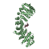
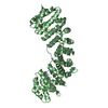
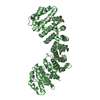
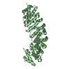
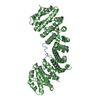
| Entry | Database: PDB / ID: 5klt | ||||||
|---|---|---|---|---|---|---|---|
| Title | Prototypical P4[M]cNLS | ||||||
 Components Components |
| ||||||
 Keywords Keywords | PROTEIN TRANSPORT/Viral protein / Importin alpha / Complex / nuclear localisation signal / PROTEIN TRANSPORT-Viral protein complex | ||||||
| Function / homology |  Function and homology information Function and homology informationSensing of DNA Double Strand Breaks / regulation of transcription by glucose / entry of viral genome into host nucleus through nuclear pore complex via importin / positive regulation of viral life cycle / NLS-dependent protein nuclear import complex / postsynapse to nucleus signaling pathway / nuclear import signal receptor activity / nuclear localization sequence binding / NLS-bearing protein import into nucleus / non-canonical NF-kappaB signal transduction ...Sensing of DNA Double Strand Breaks / regulation of transcription by glucose / entry of viral genome into host nucleus through nuclear pore complex via importin / positive regulation of viral life cycle / NLS-dependent protein nuclear import complex / postsynapse to nucleus signaling pathway / nuclear import signal receptor activity / nuclear localization sequence binding / NLS-bearing protein import into nucleus / non-canonical NF-kappaB signal transduction / positive regulation of type I interferon production / histone deacetylase binding / cytoplasmic stress granule / protein import into nucleus / host cell / nuclear membrane / DNA-binding transcription factor binding / postsynaptic density / positive regulation of DNA-templated transcription / glutamatergic synapse / nucleoplasm / nucleus / cytoplasm / cytosol Similarity search - Function | ||||||
| Biological species |  synthetic construct (others) | ||||||
| Method |  X-RAY DIFFRACTION / SYNCHROTRON / MOLECULAR REPLACEMENT / Resolution: 2.6 Å X-RAY DIFFRACTION / SYNCHROTRON / MOLECULAR REPLACEMENT / Resolution: 2.6 Å | ||||||
 Authors Authors | Smith, K.M. / Forwood, J.K. | ||||||
 Citation Citation | Journal: Biochim Biophys Acta Mol Cell Res / Year: 2018 Title: Contribution of the residue at position 4 within classical nuclear localization signals to modulating interaction with importins and nuclear targeting. Authors: Smith, K.M. / Di Antonio, V. / Bellucci, L. / Thomas, D.R. / Caporuscio, F. / Ciccarese, F. / Ghassabian, H. / Wagstaff, K.M. / Forwood, J.K. / Jans, D.A. / Palu, G. / Alvisi, G. | ||||||
| History |
|
- Structure visualization
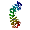
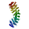
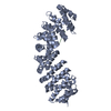
| Structure viewer | Molecule: MolmilJmol/JSmol |
|---|
- Downloads & links
-Download
| PDBx/mmCIF format | 5klt.cif.gz | 170.6 KB | Display | PDBx/mmCIF format |
|---|---|---|---|---|
| PDB format | pdb5klt.ent.gz | 135.1 KB | Display | PDB format |
| PDBx/mmJSON format | 5klt.json.gz | Tree view | PDBx/mmJSON format | |
| Others |  Other downloads Other downloads |
-Validation report
| Arichive directory | https://data.pdbj.org/pub/pdb/validation_reports/kl/5kltftp://data.pdbj.org/pub/pdb/validation_reports/kl/5klt | HTTPS FTP |
|---|
-Related structure data
| Related structure data |  5klrC  3ul1S C: citing same article ( S: Starting model for refinement |
|---|---|
| Similar structure data |


-Links
 PDBj
PDBj


- Assembly
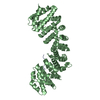
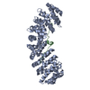
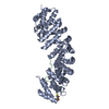
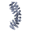
| Deposited unit | 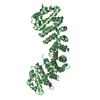
| ||||||||
|---|---|---|---|---|---|---|---|---|---|
| 1 |
| ||||||||
| Unit cell |
|
-Components
| #1: Protein | Mass: 55268.473 Da / Num. of mol.: 1 / Fragment: UNp residues 70-529 Source method: isolated from a genetically manipulated source Source: (gene. exp.)  |
|---|---|
| #2: Protein/peptide | Mass: 1696.084 Da / Num. of mol.: 1 Source method: isolated from a genetically manipulated source Source: (gene. exp.) synthetic construct (others) / Production host: |
-Experimental details
-Experiment
| Experiment | Method: X-RAY DIFFRACTION / Number of used crystals: 1 |
|---|
- Sample preparation
| Crystal | Density Matthews: 3.09 Å3/Da / Density % sol: 60.24 % |
|---|---|
| Crystal grow | Temperature: 296 K / Method: vapor diffusion, hanging drop / pH: 6 / Details: 1M Sodium citrate pH6, 10mM DTT |
-Data collection
| Diffraction | Mean temperature: 100 K | |||||||||||||||
|---|---|---|---|---|---|---|---|---|---|---|---|---|---|---|---|---|
| Diffraction source | Source: SYNCHROTRON / Site: Australian Synchrotron  / Beamline: MX1 / Wavelength: 0.9537 Å / Beamline: MX1 / Wavelength: 0.9537 Å | |||||||||||||||
| Detector | Type: ADSC QUANTUM 210r / Detector: CCD / Date: Sep 26, 2015 | |||||||||||||||
| Radiation | Protocol: SINGLE WAVELENGTH / Monochromatic (M) / Laue (L): M / Scattering type: x-ray | |||||||||||||||
| Radiation wavelength | Wavelength: 0.9537 Å / Relative weight: 1 | |||||||||||||||
| Reflection | Resolution: 2.6→26.7 Å / Num. obs: 22383 / % possible obs: 99.9 % / Redundancy: 14.1 % / Biso Wilson estimate: 34.34 Å2 / CC1/2: 0.992 / Rmerge(I) obs: 0.3 / Net I/σ(I): 9.3 | |||||||||||||||
| Reflection shell |
|
- Processing
| Software |
| ||||||||||||||||||||||||||||||||||||||||||||||||||||||
|---|---|---|---|---|---|---|---|---|---|---|---|---|---|---|---|---|---|---|---|---|---|---|---|---|---|---|---|---|---|---|---|---|---|---|---|---|---|---|---|---|---|---|---|---|---|---|---|---|---|---|---|---|---|---|---|
| Refinement | Method to determine structure: MOLECULAR REPLACEMENT Starting model: 3UL1 Resolution: 2.6→26.698 Å / SU ML: 0.32 / Cross valid method: FREE R-VALUE / σ(F): 1.34 / Phase error: 23.18
| ||||||||||||||||||||||||||||||||||||||||||||||||||||||
| Solvent computation | Shrinkage radii: 0.9 Å / VDW probe radii: 1.11 Å | ||||||||||||||||||||||||||||||||||||||||||||||||||||||
| Displacement parameters | Biso max: 132.09 Å2 / Biso mean: 44.5811 Å2 / Biso min: 19.17 Å2 | ||||||||||||||||||||||||||||||||||||||||||||||||||||||
| Refinement step | Cycle: final / Resolution: 2.6→26.698 Å
| ||||||||||||||||||||||||||||||||||||||||||||||||||||||
| Refine LS restraints |
| ||||||||||||||||||||||||||||||||||||||||||||||||||||||
| LS refinement shell | Refine-ID: X-RAY DIFFRACTION / Total num. of bins used: 8 / % reflection obs: 100 %
|
