 Movie
Movie Controller

[English] 日本語
 Yorodumi
- PDB-6iu7: Crystal structure of importin-alpha1 bound to the 53BP1 nuclear l... -
+ Open data

- Basic information
| Entry | Database: PDB / ID: 6iu7 | ||||||
|---|---|---|---|---|---|---|---|
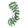
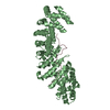
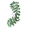
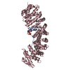
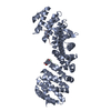
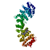
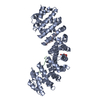
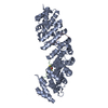
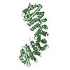
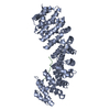
| Title | Crystal structure of importin-alpha1 bound to the 53BP1 nuclear localization signal (wild-type) | ||||||
 Components Components |
| ||||||
 Keywords Keywords | TRANSPORT PROTEIN/PROTEIN BINDING / importin / NLS / TRANSPORT PROTEIN / TRANSPORT PROTEIN-PROTEIN BINDING complex | ||||||
| Function / homology |  Function and homology information Function and homology informationhistone H4K20me methyltransferase activity / positive regulation of isotype switching / Sensing of DNA Double Strand Breaks / ubiquitin-modified histone reader activity / regulation of transcription by glucose / histone H4K20me2 reader activity / entry of viral genome into host nucleus through nuclear pore complex via importin / positive regulation of viral life cycle / NLS-dependent protein nuclear import complex / cellular response to X-ray ...histone H4K20me methyltransferase activity / positive regulation of isotype switching / Sensing of DNA Double Strand Breaks / ubiquitin-modified histone reader activity / regulation of transcription by glucose / histone H4K20me2 reader activity / entry of viral genome into host nucleus through nuclear pore complex via importin / positive regulation of viral life cycle / NLS-dependent protein nuclear import complex / cellular response to X-ray / postsynapse to nucleus signaling pathway / double-strand break repair via classical nonhomologous end joining / protein localization to site of double-strand break / DNA repair complex / nuclear import signal receptor activity / nuclear localization sequence binding / NLS-bearing protein import into nucleus / non-canonical NF-kappaB signal transduction / telomeric repeat DNA binding / SUMOylation of transcription factors / histone reader activity / positive regulation of intrinsic apoptotic signaling pathway by p53 class mediator / negative regulation of double-strand break repair via homologous recombination / positive regulation of type I interferon production / DNA damage checkpoint signaling / replication fork / transcription coregulator activity / Nonhomologous End-Joining (NHEJ) / protein homooligomerization / G2/M DNA damage checkpoint / protein import into nucleus / double-strand break repair via nonhomologous end joining / kinetochore / histone deacetylase binding / cytoplasmic stress granule / p53 binding / host cell / Recruitment and ATM-mediated phosphorylation of repair and signaling proteins at DNA double strand breaks / site of double-strand break / Processing of DNA double-strand break ends / nuclear membrane / histone binding / DNA-binding transcription factor binding / damaged DNA binding / RNA polymerase II-specific DNA-binding transcription factor binding / transcription coactivator activity / chromosome, telomeric region / postsynaptic density / nuclear body / DNA damage response / positive regulation of DNA-templated transcription / glutamatergic synapse / positive regulation of transcription by RNA polymerase II / nucleoplasm / nucleus / cytosol / cytoplasm Similarity search - Function | ||||||
| Biological species |   Homo sapiens (human) Homo sapiens (human) | ||||||
| Method |  X-RAY DIFFRACTION / SYNCHROTRON / MOLECULAR REPLACEMENT / Resolution: 1.9 Å X-RAY DIFFRACTION / SYNCHROTRON / MOLECULAR REPLACEMENT / Resolution: 1.9 Å | ||||||
 Authors Authors | Matsuura, Y. | ||||||
 Citation Citation | Journal: Biochem. Biophys. Res. Commun. / Year: 2019 Title: Structural and biochemical characterization of the recognition of the 53BP1 nuclear localization signal by importin-alpha. Authors: Matsuura, Y. | ||||||
| History |
|
- Structure visualization
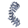
| Structure viewer | Molecule: MolmilJmol/JSmol |
|---|
- Downloads & links
-Download
| PDBx/mmCIF format | 6iu7.cif.gz | 191.9 KB | Display | PDBx/mmCIF format |
|---|---|---|---|---|
| PDB format | pdb6iu7.ent.gz | 150.8 KB | Display | PDB format |
| PDBx/mmJSON format | 6iu7.json.gz | Tree view | PDBx/mmJSON format | |
| Others |  Other downloads Other downloads |
-Validation report
| Arichive directory | https://data.pdbj.org/pub/pdb/validation_reports/iu/6iu7ftp://data.pdbj.org/pub/pdb/validation_reports/iu/6iu7 | HTTPS FTP |
|---|
-Related structure data
| Related structure data |  6iuaC  5x8nS C: citing same article ( S: Starting model for refinement |
|---|---|
| Similar structure data |


-Links
 PDBj
PDBj


- Assembly
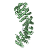
| Deposited unit | 
| ||||||||
|---|---|---|---|---|---|---|---|---|---|
| 1 |
| ||||||||
| Unit cell |
|
-Components
| #1: Protein | Mass: 46242.957 Da / Num. of mol.: 1 Source method: isolated from a genetically manipulated source Source: (gene. exp.)  |
|---|---|
| #2: Protein/peptide | Mass: 2508.878 Da / Num. of mol.: 1 Source method: isolated from a genetically manipulated source Source: (gene. exp.) Homo sapiens (human) / Gene: TP53BP1 / Production host: |
| #3: Water | ChemComp-HOH /  Mass: 18.015 Da / Num. of mol.: 316 / Source method: isolated from a natural source / Formula: H2O Mass: 18.015 Da / Num. of mol.: 316 / Source method: isolated from a natural source / Formula: H2O |
-Experimental details
-Experiment
| Experiment | Method: X-RAY DIFFRACTION / Number of used crystals: 1 |
|---|
- Sample preparation
| Crystal | Density Matthews: 3.55 Å3/Da / Density % sol: 65.38 % |
|---|---|
| Crystal grow | Temperature: 293 K / Method: vapor diffusion, hanging drop / pH: 6.7 / Details: sodium citrate, HEPES, DTT |
-Data collection
| Diffraction | Mean temperature: 95 K / Serial crystal experiment: N |
|---|---|
| Diffraction source | Source: SYNCHROTRON / Site: Photon Factory  / Beamline: BL-17A / Wavelength: 0.98 Å / Beamline: BL-17A / Wavelength: 0.98 Å |
| Detector | Type: DECTRIS EIGER X 16M / Detector: PIXEL / Date: Jun 23, 2018 |
| Radiation | Protocol: SINGLE WAVELENGTH / Monochromatic (M) / Laue (L): M / Scattering type: x-ray |
| Radiation wavelength | Wavelength: 0.98 Å / Relative weight: 1 |
| Reflection | Resolution: 1.9→32.973 Å / Num. obs: 54893 / % possible obs: 99.2 % / Redundancy: 3.4 % / Biso Wilson estimate: 26.77 Å2 / CC1/2: 0.996 / Rmerge(I) obs: 0.072 / Rpim(I) all: 0.045 / Rrim(I) all: 0.086 / Net I/σ(I): 8.9 |
| Reflection shell | Resolution: 1.9→1.94 Å / Redundancy: 3.5 % / Rmerge(I) obs: 0.674 / Num. unique obs: 3481 / CC1/2: 0.71 / Rpim(I) all: 0.408 / Rrim(I) all: 0.791 / % possible all: 99 |
- Processing
| Software |
| ||||||||||||||||||||||||||||||||||||||||||||||||||||||||||||||||||||||||||||||||||||||||||||||||||||||||||||||||||||||||||||||||||||||||||||||||||||||||||||||||||||||||||||||||||||||||||||||||||||||||||||||||||||||||||||||||||||||||||||||||||||||||||||||||||||||||||||||||||||||||||||||||||||||||||||||||||||||||||||||||||||||||||||||||||||||||||||||
|---|---|---|---|---|---|---|---|---|---|---|---|---|---|---|---|---|---|---|---|---|---|---|---|---|---|---|---|---|---|---|---|---|---|---|---|---|---|---|---|---|---|---|---|---|---|---|---|---|---|---|---|---|---|---|---|---|---|---|---|---|---|---|---|---|---|---|---|---|---|---|---|---|---|---|---|---|---|---|---|---|---|---|---|---|---|---|---|---|---|---|---|---|---|---|---|---|---|---|---|---|---|---|---|---|---|---|---|---|---|---|---|---|---|---|---|---|---|---|---|---|---|---|---|---|---|---|---|---|---|---|---|---|---|---|---|---|---|---|---|---|---|---|---|---|---|---|---|---|---|---|---|---|---|---|---|---|---|---|---|---|---|---|---|---|---|---|---|---|---|---|---|---|---|---|---|---|---|---|---|---|---|---|---|---|---|---|---|---|---|---|---|---|---|---|---|---|---|---|---|---|---|---|---|---|---|---|---|---|---|---|---|---|---|---|---|---|---|---|---|---|---|---|---|---|---|---|---|---|---|---|---|---|---|---|---|---|---|---|---|---|---|---|---|---|---|---|---|---|---|---|---|---|---|---|---|---|---|---|---|---|---|---|---|---|---|---|---|---|---|---|---|---|---|---|---|---|---|---|---|---|---|---|---|---|---|---|---|---|---|---|---|---|---|---|---|---|---|---|---|---|---|---|---|---|---|---|---|---|---|---|---|---|---|---|---|---|---|---|---|---|---|---|---|---|---|---|---|---|---|---|---|---|---|---|---|---|---|---|---|---|---|---|---|---|---|---|---|---|---|---|---|
| Refinement | Method to determine structure: MOLECULAR REPLACEMENT Starting model: 5X8N Resolution: 1.9→32.973 Å / SU ML: 0.16 / Cross valid method: THROUGHOUT / σ(F): 1.35 / Phase error: 16.51
| ||||||||||||||||||||||||||||||||||||||||||||||||||||||||||||||||||||||||||||||||||||||||||||||||||||||||||||||||||||||||||||||||||||||||||||||||||||||||||||||||||||||||||||||||||||||||||||||||||||||||||||||||||||||||||||||||||||||||||||||||||||||||||||||||||||||||||||||||||||||||||||||||||||||||||||||||||||||||||||||||||||||||||||||||||||||||||||||
| Solvent computation | Shrinkage radii: 0.9 Å / VDW probe radii: 1.11 Å | ||||||||||||||||||||||||||||||||||||||||||||||||||||||||||||||||||||||||||||||||||||||||||||||||||||||||||||||||||||||||||||||||||||||||||||||||||||||||||||||||||||||||||||||||||||||||||||||||||||||||||||||||||||||||||||||||||||||||||||||||||||||||||||||||||||||||||||||||||||||||||||||||||||||||||||||||||||||||||||||||||||||||||||||||||||||||||||||
| Displacement parameters | Biso max: 118.42 Å2 / Biso mean: 37.0533 Å2 / Biso min: 14.81 Å2 | ||||||||||||||||||||||||||||||||||||||||||||||||||||||||||||||||||||||||||||||||||||||||||||||||||||||||||||||||||||||||||||||||||||||||||||||||||||||||||||||||||||||||||||||||||||||||||||||||||||||||||||||||||||||||||||||||||||||||||||||||||||||||||||||||||||||||||||||||||||||||||||||||||||||||||||||||||||||||||||||||||||||||||||||||||||||||||||||
| Refinement step | Cycle: final / Resolution: 1.9→32.973 Å
| ||||||||||||||||||||||||||||||||||||||||||||||||||||||||||||||||||||||||||||||||||||||||||||||||||||||||||||||||||||||||||||||||||||||||||||||||||||||||||||||||||||||||||||||||||||||||||||||||||||||||||||||||||||||||||||||||||||||||||||||||||||||||||||||||||||||||||||||||||||||||||||||||||||||||||||||||||||||||||||||||||||||||||||||||||||||||||||||
| LS refinement shell | Refine-ID: X-RAY DIFFRACTION / Rfactor Rfree error: 0 / Total num. of bins used: 20
| ||||||||||||||||||||||||||||||||||||||||||||||||||||||||||||||||||||||||||||||||||||||||||||||||||||||||||||||||||||||||||||||||||||||||||||||||||||||||||||||||||||||||||||||||||||||||||||||||||||||||||||||||||||||||||||||||||||||||||||||||||||||||||||||||||||||||||||||||||||||||||||||||||||||||||||||||||||||||||||||||||||||||||||||||||||||||||||||
| Refinement TLS params. | Method: refined / Refine-ID: X-RAY DIFFRACTION
| ||||||||||||||||||||||||||||||||||||||||||||||||||||||||||||||||||||||||||||||||||||||||||||||||||||||||||||||||||||||||||||||||||||||||||||||||||||||||||||||||||||||||||||||||||||||||||||||||||||||||||||||||||||||||||||||||||||||||||||||||||||||||||||||||||||||||||||||||||||||||||||||||||||||||||||||||||||||||||||||||||||||||||||||||||||||||||||||
| Refinement TLS group |
|
