[English] 日本語
Yorodumi
- PDB-5x8n: Crystal structure of mouse importin-alpha1 bound to the nuclear l... -

+
Open data


ID or keywords:

Loading...

-
Basic information

Entry
Database: PDB / ID: 5x8n
Title
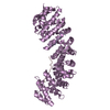
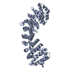
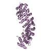
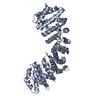
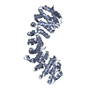
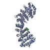
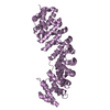
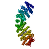
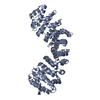
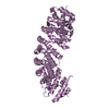
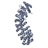
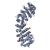
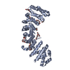
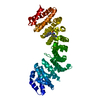
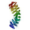
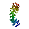
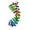
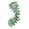
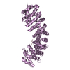
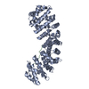
Crystal structure of mouse importin-alpha1 bound to the nuclear localization signal of Epstein-Barr virus EBNA-LP protein
Components
  • Epstein-Barr nuclear antigen leader protein
  • Importin subunit alpha-1
KeywordsPROTEIN TRANSPORT/TRANSCRIPTION / importin / NLS / EBNA-LP / Epstein-Barr virus / PROTEIN TRANSPORT-TRANSCRIPTION complex
Function / homology
Function and homology information


host cell PML body / DNA-templated viral transcription / Sensing of DNA Double Strand Breaks / entry of viral genome into host nucleus through nuclear pore complex via importin / positive regulation of viral life cycle / NLS-dependent protein nuclear import complex / postsynapse to nucleus signaling pathway / host cell / symbiont-mediated disruption of host cell PML body / nuclear import signal receptor activity ...host cell PML body / DNA-templated viral transcription / Sensing of DNA Double Strand Breaks / entry of viral genome into host nucleus through nuclear pore complex via importin / positive regulation of viral life cycle / NLS-dependent protein nuclear import complex / postsynapse to nucleus signaling pathway / host cell / symbiont-mediated disruption of host cell PML body / nuclear import signal receptor activity / nuclear localization sequence binding / NLS-bearing protein import into nucleus / cytoplasmic stress granule / protein import into nucleus / histone deacetylase binding / nuclear membrane / DNA-binding transcription factor binding / postsynaptic density / transcription coactivator activity / glutamatergic synapse / host cell nucleus / nucleoplasm / nucleus / cytosol
Similarity search - Function
Herpesvirus leader protein / Herpesvirus leader protein / Importin subunit alpha / Atypical Arm repeat / Importin-alpha, importin-beta-binding domain superfamily / Importin beta binding domain / Atypical Arm repeat / Importin-alpha, importin-beta-binding domain / IBB domain profile. / Armadillo/plakoglobin ARM repeat profile. ...Herpesvirus leader protein / Herpesvirus leader protein / Importin subunit alpha / Atypical Arm repeat / Importin-alpha, importin-beta-binding domain superfamily / Importin beta binding domain / Atypical Arm repeat / Importin-alpha, importin-beta-binding domain / IBB domain profile. / Armadillo/plakoglobin ARM repeat profile. / Armadillo/beta-catenin-like repeat / Armadillo/beta-catenin-like repeats / Armadillo / Leucine-rich Repeat Variant / Leucine-rich Repeat Variant / Armadillo-like helical / Alpha Horseshoe / Armadillo-type fold / Mainly Alpha
Similarity search - Domain/homology
Importin subunit alpha-1 / Epstein-Barr nuclear antigen leader protein
Similarity search - Component
Biological speciesMus musculus (house mouse)
Human herpesvirus 4 (Epstein-Barr virus)
MethodX-RAY DIFFRACTION / SYNCHROTRON / MOLECULAR REPLACEMENT / molecular replacement / Resolution: 2.15 Å
AuthorsNakada, R. / Matsuura, Y.
CitationJournal: Protein Sci. / Year: 2017
Title: Crystal structure of importin-alpha bound to the nuclear localization signal of Epstein-Barr virus EBNA-LP protein
Authors: Nakada, R. / Matsuura, Y.
History
DepositionMar 3, 2017Deposition site: PDBJ / Processing site: PDBJ
Revision 1.0Apr 19, 2017Provider: repository / Type: Initial release
Revision 1.1Jun 7, 2017Group: Database references
Revision 1.2Nov 22, 2023Group: Data collection / Database references / Refinement description
Category: chem_comp_atom / chem_comp_bond ...chem_comp_atom / chem_comp_bond / database_2 / pdbx_initial_refinement_model
Item: _database_2.pdbx_DOI / _database_2.pdbx_database_accession

-
Structure visualization

Structure viewerMolecule:
MolmilJmol/JSmol

Downloads & links

-
Assembly

Deposited unit
A: Importin subunit alpha-1
B: Epstein-Barr nuclear antigen leader protein


Theoretical massNumber of molelcules
Total (without water)52,5942
Polymers52,5942
Non-polymers00
Water2,216123
1


  • Idetical with deposited unit
  • defined by author&software
TypeNameSymmetry operationNumber
identity operation1_555x,y,z1
Buried area1600 Å2
ΔGint3 kcal/mol
Surface area18210 Å2
MethodPISA
Unit cell
Length a, b, c (Å)78.270, 90.290, 98.170
Angle α, β, γ (deg.)90.000, 90.000, 90.000
Int Tables number19
Space group name H-MP212121

-
Components

#1: Protein Importin subunit alpha-1 / / Importin alpha P1 / Karyopherin subunit alpha-2 / Pendulin / Pore targeting complex 58 kDa subunit ...Importin alpha P1 / Karyopherin subunit alpha-2 / Pendulin / Pore targeting complex 58 kDa subunit / PTAC58 / RAG cohort protein 1 / SRP1-alpha


Mass: 49886.633 Da / Num. of mol.: 1 / Fragment: UNP residues 70-529
Source method: isolated from a genetically manipulated source
Source: (gene. exp.) Mus musculus (house mouse) / Gene: Kpna2, Rch1 / Production host: Escherichia coli (E. coli) / References: UniProt: P52293
#2: Protein/peptide Epstein-Barr nuclear antigen leader protein / EBV nuclear antigen leader protein / Epstein-Barr nuclear antigen 5 / EBV nuclear antigen 5


Mass: 2707.118 Da / Num. of mol.: 1 / Fragment: UNP residues 29-50 / Source method: obtained synthetically
Source: (synth.) Human herpesvirus 4 (strain B95-8) (Epstein-Barr virus (strain B95-8))
References: UniProt: Q8AZK7
#3: Water ChemComp-HOH / water / Water


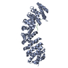
Mass: 18.015 Da / Num. of mol.: 123 / Source method: isolated from a natural source / Formula: H2O

-
Experimental details

-
Experiment

ExperimentMethod: X-RAY DIFFRACTION / Number of used crystals: 1

-
Sample preparation

CrystalDensity Matthews: 3.3 Å3/Da / Density % sol: 62.7 %
Crystal growTemperature: 293 K / Method: vapor diffusion, hanging drop / pH: 7 / Details: MES, sodium citrate, DTT

-
Data collection

DiffractionMean temperature: 95 K
Diffraction sourceSource: SYNCHROTRON / Site: Photon Factory / Beamline: BL-17A / Wavelength: 0.98 Å
DetectorType: DECTRIS PILATUS3 S 6M / Detector: PIXEL / Date: Feb 12, 2017
RadiationProtocol: SINGLE WAVELENGTH / Monochromatic (M) / Laue (L): M / Scattering type: x-ray
Radiation wavelengthWavelength: 0.98 Å / Relative weight: 1
ReflectionResolution: 2.15→30.77 Å / Num. obs: 38548 / % possible obs: 100 % / Redundancy: 5.9 % / Biso Wilson estimate: 38.14 Å2 / CC1/2: 0.998 / Rmerge(I) obs: 0.086 / Rpim(I) all: 0.039 / Rrim(I) all: 0.095 / Net I/σ(I): 13.1 / Num. measured all: 227759 / Scaling rejects: 138
Reflection shell

Diffraction-ID: 1

Resolution (Å)Redundancy (%)Rmerge(I) obsCC1/2Rpim(I) allRrim(I) all% possible all
2.15-2.225.80.9640.680.4361.06100
8.86-30.7750.0340.9990.0170.03898.1

-
Phasing

PhasingMethod: molecular replacement

-
Processing

Software
NameVersionClassification
MOSFLMdata collection
Aimless0.5.31data scaling
MOLREPphasing
PHENIX1.11.1_2575refinement
PDB_EXTRACT3.22data extraction
Cootmodel building
Aimlessdata processing
MOSFLMdata reduction
RefinementMethod to determine structure: MOLECULAR REPLACEMENT
Starting model: 5WUM
Resolution: 2.15→30.765 Å / SU ML: 0.23 / Cross valid method: FREE R-VALUE / σ(F): 1.33 / Phase error: 20.99
RfactorNum. reflection% reflection
Rfree0.2099 1896 4.93 %
Rwork0.1852 --
obs0.1865 38484 99.94 %
Solvent computationShrinkage radii: 0.9 Å / VDW probe radii: 1.11 Å
Displacement parametersBiso max: 157.49 Å2 / Biso mean: 48.2742 Å2 / Biso min: 22.18 Å2
Refinement stepCycle: final / Resolution: 2.15→30.765 Å
ProteinNucleic acidLigandSolventTotal
Num. atoms3316 0 0 123 3439
Biso mean---48.63 -
Num. residues----433
Refine LS restraints
Refine-IDTypeDev idealNumber
X-RAY DIFFRACTIONf_bond_d0.0033374
X-RAY DIFFRACTIONf_angle_d0.6564593
X-RAY DIFFRACTIONf_chiral_restr0.04554
X-RAY DIFFRACTIONf_plane_restr0.004587
X-RAY DIFFRACTIONf_dihedral_angle_d2.632056
LS refinement shell

Refine-ID: X-RAY DIFFRACTION / Rfactor Rfree error: 0 / Total num. of bins used: 14 / % reflection obs: 100 %

Resolution (Å)Rfactor RfreeNum. reflection RfreeRfactor RworkNum. reflection RworkNum. reflection all
2.15-2.20380.31161240.271625732697
2.2038-2.26330.28511380.243925652703
2.2633-2.32990.27471250.232225822707
2.3299-2.40510.26941320.226125752707
2.4051-2.4910.24761470.203425792726
2.491-2.59070.24021420.207725712713
2.5907-2.70850.24361360.198226012737
2.7085-2.85120.22961300.184526002730
2.8512-3.02970.21271330.194426182751
3.0297-3.26340.23031190.195226182737
3.2634-3.59140.18481190.187126462765
3.5914-4.110.17851500.165226292779
4.11-5.17420.17851440.1626532797
5.1742-30.76820.20331570.169927782935

+
About Yorodumi

-
News

-
Feb 9, 2022. New format data for meta-information of EMDB entries

New format data for meta-information of EMDB entries

  • Version 3 of the EMDB header file is now the official format.
  • The previous official version 1.9 will be removed from the archive.

Related info.:EMDB header

External links:wwPDB to switch to version 3 of the EMDB data model

-
Aug 12, 2020. Covid-19 info

Covid-19 info

URL: https://pdbj.org/emnavi/covid19.php

New page: Covid-19 featured information page in EM Navigator.

Related info.:Covid-19 info / Mar 5, 2020. Novel coronavirus structure data

+
Mar 5, 2020. Novel coronavirus structure data

Novel coronavirus structure data

Related info.:Yorodumi Speices / Aug 12, 2020. Covid-19 info

External links:COVID-19 featured content - PDBj / Molecule of the Month (242):Coronavirus Proteases

+
Jan 31, 2019. EMDB accession codes are about to change! (news from PDBe EMDB page)

EMDB accession codes are about to change! (news from PDBe EMDB page)

  • The allocation of 4 digits for EMDB accession codes will soon come to an end. Whilst these codes will remain in use, new EMDB accession codes will include an additional digit and will expand incrementally as the available range of codes is exhausted. The current 4-digit format prefixed with “EMD-” (i.e. EMD-XXXX) will advance to a 5-digit format (i.e. EMD-XXXXX), and so on. It is currently estimated that the 4-digit codes will be depleted around Spring 2019, at which point the 5-digit format will come into force.
  • The EM Navigator/Yorodumi systems omit the EMD- prefix.

Related info.:Q: What is EMD? / ID/Accession-code notation in Yorodumi/EM Navigator

External links:EMDB Accession Codes are Changing Soon! / Contact to PDBj

+
Jul 12, 2017. Major update of PDB

Major update of PDB

  • wwPDB released updated PDB data conforming to the new PDBx/mmCIF dictionary.
  • This is a major update changing the version number from 4 to 5, and with Remediation, in which all the entries are updated.
  • In this update, many items about electron microscopy experimental information are reorganized (e.g. em_software).
  • Now, EM Navigator and Yorodumi are based on the updated data.

External links:wwPDB Remediation / Enriched Model Files Conforming to OneDep Data Standards Now Available in the PDB FTP Archive

-
Yorodumi

Thousand views of thousand structures

  • Yorodumi is a browser for structure data from EMDB, PDB, SASBDB, etc.
  • This page is also the successor to EM Navigator detail page, and also detail information page/front-end page for Omokage search.
  • The word "yorodu" (or yorozu) is an old Japanese word meaning "ten thousand". "mi" (miru) is to see.

Related info.:EMDB / PDB / SASBDB / Comparison of 3 databanks / Yorodumi Search / Aug 31, 2016. New EM Navigator & Yorodumi / Yorodumi Papers / Jmol/JSmol / Function and homology information / Changes in new EM Navigator and Yorodumi

Read more