 Movie
Movie Controller

[English] 日本語
 Yorodumi
- PDB-7l04: Crystal Structure of Adeno-Associated Virus Porcine Origin capsid... -
+ Open data

- Basic information
| Entry | Database: PDB / ID: 7l04 | |||||||||
|---|---|---|---|---|---|---|---|---|---|---|
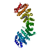
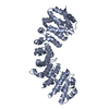
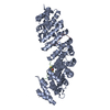
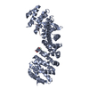
| Title | Crystal Structure of Adeno-Associated Virus Porcine Origin capsid protein in complex with Importin-alpha 2 | |||||||||
 Components Components |
| |||||||||
 Keywords Keywords | TRANSPORT PROTEIN / Complex / transport / Nuclear Localization | |||||||||
| Function / homology |  Function and homology information Function and homology informationSensing of DNA Double Strand Breaks / regulation of transcription by glucose / entry of viral genome into host nucleus through nuclear pore complex via importin / positive regulation of viral life cycle / NLS-dependent protein nuclear import complex / postsynapse to nucleus signaling pathway / NLS-bearing protein import into nucleus / nuclear import signal receptor activity / nuclear localization sequence binding / non-canonical NF-kappaB signal transduction ...Sensing of DNA Double Strand Breaks / regulation of transcription by glucose / entry of viral genome into host nucleus through nuclear pore complex via importin / positive regulation of viral life cycle / NLS-dependent protein nuclear import complex / postsynapse to nucleus signaling pathway / NLS-bearing protein import into nucleus / nuclear import signal receptor activity / nuclear localization sequence binding / non-canonical NF-kappaB signal transduction / T=1 icosahedral viral capsid / positive regulation of type I interferon production / protein import into nucleus / histone deacetylase binding / cytoplasmic stress granule / host cell / nuclear membrane / DNA-binding transcription factor binding / postsynaptic density / positive regulation of DNA-templated transcription / glutamatergic synapse / structural molecule activity / nucleoplasm / nucleus / cytosol / cytoplasm Similarity search - Function | |||||||||
| Biological species |  Adeno-associated virus - Po1 Adeno-associated virus - Po1 | |||||||||
| Method |  X-RAY DIFFRACTION / SYNCHROTRON / MOLECULAR REPLACEMENT / Resolution: 2.26 Å X-RAY DIFFRACTION / SYNCHROTRON / MOLECULAR REPLACEMENT / Resolution: 2.26 Å | |||||||||
 Authors Authors | Hoad, M. / Forwood, J. | |||||||||
 Citation Citation | Journal: To Be Published Title: Crystal Structure of Adeno-Associated Virus Porcine Origin capsid protein in complex with Importin-alpha 2 Authors: Hoad, M. / Forwood, J. | |||||||||
| History |
|
- Structure visualization
| Structure viewer | Molecule: MolmilJmol/JSmol |
|---|
- Downloads & links
-Download
| PDBx/mmCIF format | 7l04.cif.gz | 209.5 KB | Display | PDBx/mmCIF format |
|---|---|---|---|---|
| PDB format | pdb7l04.ent.gz | 134.8 KB | Display | PDB format |
| PDBx/mmJSON format | 7l04.json.gz | Tree view | PDBx/mmJSON format | |
| Others |  Other downloads Other downloads |
-Validation report
| Arichive directory | https://data.pdbj.org/pub/pdb/validation_reports/l0/7l04ftp://data.pdbj.org/pub/pdb/validation_reports/l0/7l04 | HTTPS FTP |
|---|
-Related structure data
| Related structure data |  6bw0S S: Starting model for refinement |
|---|---|
| Similar structure data |

-Links
 PDBj
PDBj


- Assembly
| Deposited unit | 
| ||||||||||||
|---|---|---|---|---|---|---|---|---|---|---|---|---|---|
| 1 |
| ||||||||||||
| Unit cell |
|
-Components
| #1: Protein/peptide | Mass: 1265.566 Da / Num. of mol.: 1 Source method: isolated from a genetically manipulated source Source: (gene. exp.) Adeno-associated virus - Po1 / Production host:  |
|---|---|
| #2: Protein | Mass: 55268.473 Da / Num. of mol.: 1 Source method: isolated from a genetically manipulated source Source: (gene. exp.) |
| #3: Water | ChemComp-HOH /  Mass: 18.015 Da / Num. of mol.: 54 / Source method: isolated from a natural source / Formula: H2O Mass: 18.015 Da / Num. of mol.: 54 / Source method: isolated from a natural source / Formula: H2O |
-Experimental details
-Experiment
| Experiment | Method: X-RAY DIFFRACTION / Number of used crystals: 1 |
|---|
- Sample preparation
| Crystal | Density Matthews: 3.13 Å3/Da / Density % sol: 60.68 % |
|---|---|
| Crystal grow | Temperature: 298 K / Method: vapor diffusion, hanging drop / pH: 6.5 / Details: 0.6M Na citrate, 0.01M DTT, 0.1M HEPES pH 6.5 |
-Data collection
| Diffraction | Mean temperature: 100 K / Serial crystal experiment: N |
|---|---|
| Diffraction source | Source: SYNCHROTRON / Site: Australian Synchrotron  / Beamline: MX2 / Wavelength: 0.9537 Å / Beamline: MX2 / Wavelength: 0.9537 Å |
| Detector | Type: DECTRIS EIGER X 16M / Detector: PIXEL / Date: Oct 7, 2020 |
| Radiation | Protocol: SINGLE WAVELENGTH / Monochromatic (M) / Laue (L): M / Scattering type: x-ray |
| Radiation wavelength | Wavelength: 0.9537 Å / Relative weight: 1 |
| Reflection | Resolution: 2.26→44.67 Å / Num. obs: 33929 / % possible obs: 99.8 % / Redundancy: 6.8 % / Biso Wilson estimate: 34.4 Å2 / CC1/2: 0.996 / Rmerge(I) obs: 0.124 / Net I/σ(I): 10.1 |
| Reflection shell | Resolution: 2.26→2.33 Å / Rmerge(I) obs: 0.888 / Num. unique obs: 3034 / CC1/2: 0.759 |
- Processing
| Software |
| |||||||||||||||||||||||||||||||||||||||||||||||||||||||||||||||||||||||||||||||||||||||||||
|---|---|---|---|---|---|---|---|---|---|---|---|---|---|---|---|---|---|---|---|---|---|---|---|---|---|---|---|---|---|---|---|---|---|---|---|---|---|---|---|---|---|---|---|---|---|---|---|---|---|---|---|---|---|---|---|---|---|---|---|---|---|---|---|---|---|---|---|---|---|---|---|---|---|---|---|---|---|---|---|---|---|---|---|---|---|---|---|---|---|---|---|---|
| Refinement | Method to determine structure: MOLECULAR REPLACEMENT Starting model: 6bw0 Resolution: 2.26→44.67 Å / SU ML: 0.278 / Cross valid method: FREE R-VALUE / σ(F): 1.35 / Phase error: 23.3607 Stereochemistry target values: GeoStd + Monomer Library + CDL v1.2
| |||||||||||||||||||||||||||||||||||||||||||||||||||||||||||||||||||||||||||||||||||||||||||
| Solvent computation | Shrinkage radii: 0.9 Å / VDW probe radii: 1.11 Å / Solvent model: FLAT BULK SOLVENT MODEL | |||||||||||||||||||||||||||||||||||||||||||||||||||||||||||||||||||||||||||||||||||||||||||
| Displacement parameters | Biso mean: 45.61 Å2 | |||||||||||||||||||||||||||||||||||||||||||||||||||||||||||||||||||||||||||||||||||||||||||
| Refinement step | Cycle: LAST / Resolution: 2.26→44.67 Å
| |||||||||||||||||||||||||||||||||||||||||||||||||||||||||||||||||||||||||||||||||||||||||||
| Refine LS restraints |
| |||||||||||||||||||||||||||||||||||||||||||||||||||||||||||||||||||||||||||||||||||||||||||
| LS refinement shell |
|
