 Movie
Movie Controller

[English] 日本語
 Yorodumi
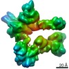
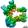
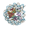
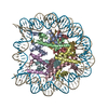
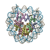
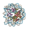
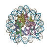
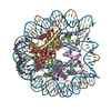
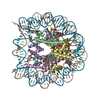
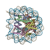
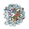
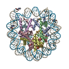
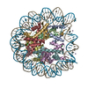
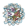
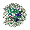
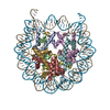
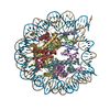
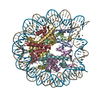
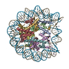
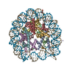
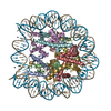
- PDB-6wz9: Bridging of double-strand DNA break activates PARP2/HPF1 to modif... -
+ Open data

- Basic information
| Entry | Database: PDB / ID: 6wz9 | |||||||||||||||||||||||||||||||||
|---|---|---|---|---|---|---|---|---|---|---|---|---|---|---|---|---|---|---|---|---|---|---|---|---|---|---|---|---|---|---|---|---|---|---|
| Title | Bridging of double-strand DNA break activates PARP2/HPF1 to modify chromatin | |||||||||||||||||||||||||||||||||
 Components Components |
| |||||||||||||||||||||||||||||||||
 Keywords Keywords | GENE REGULATION / DNA repair / PARP1 / PARP2 / HPF1 / ADP-ribosylation / chromatin / histone modifications | |||||||||||||||||||||||||||||||||
| Function / homology |  Function and homology information Function and homology informationstructural constituent of chromatin / nucleosome / heterochromatin formation / nucleosome assembly / protein heterodimerization activity / DNA binding / nucleoplasm / nucleus Similarity search - Function | |||||||||||||||||||||||||||||||||
| Biological species | synthetic construct (others) | |||||||||||||||||||||||||||||||||
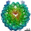
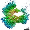
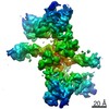
| Method | ELECTRON MICROSCOPY / single particle reconstruction / cryo EM / Resolution: 2.8 Å | |||||||||||||||||||||||||||||||||
 Authors Authors | Halic, M. / Bilokapic, S. | |||||||||||||||||||||||||||||||||
| Funding support |  United States, 1items United States, 1items
| |||||||||||||||||||||||||||||||||
 Citation Citation | Journal: Nature / Year: 2020 Title: Bridging of DNA breaks activates PARP2-HPF1 to modify chromatin. Authors: Silvija Bilokapic / Marcin J Suskiewicz / Ivan Ahel / Mario Halic /  Abstract: Breaks in DNA strands recruit the protein PARP1 and its paralogue PARP2 to modify histones and other substrates through the addition of mono- and poly(ADP-ribose) (PAR). In the DNA damage responses, ...Breaks in DNA strands recruit the protein PARP1 and its paralogue PARP2 to modify histones and other substrates through the addition of mono- and poly(ADP-ribose) (PAR). In the DNA damage responses, this post-translational modification occurs predominantly on serine residues and requires HPF1, an accessory factor that switches the amino acid specificity of PARP1 and PARP2 from aspartate or glutamate to serine. Poly(ADP) ribosylation (PARylation) is important for subsequent chromatin decompaction and provides an anchor for the recruitment of downstream signalling and repair factors to the sites of DNA breaks. Here, to understand the molecular mechanism by which PARP enzymes recognize DNA breaks within chromatin, we determined the cryo-electron-microscopic structure of human PARP2-HPF1 bound to a nucleosome. This showed that PARP2-HPF1 bridges two nucleosomes, with the broken DNA aligned in a position suitable for ligation, revealing the initial step in the repair of double-strand DNA breaks. The bridging induces structural changes in PARP2 that signal the recognition of a DNA break to the catalytic domain, which licenses HPF1 binding and PARP2 activation. Our data suggest that active PARP2 cycles through different conformational states to exchange NAD and substrate, which may enable PARP enzymes to act processively while bound to chromatin. The processes of PARP activation and the PARP catalytic cycle we describe can explain mechanisms of resistance to PARP inhibitors and will aid the development of better inhibitors as cancer treatments. | |||||||||||||||||||||||||||||||||
| History |
|
- Structure visualization
| Movie |
Movie viewer |
|---|---|
| Structure viewer | Molecule: MolmilJmol/JSmol |
- Downloads & links
-Download
| PDBx/mmCIF format | 6wz9.cif.gz | 283.4 KB | Display | PDBx/mmCIF format |
|---|---|---|---|---|
| PDB format | pdb6wz9.ent.gz | 212.6 KB | Display | PDB format |
| PDBx/mmJSON format | 6wz9.json.gz | Tree view | PDBx/mmJSON format | |
| Others |  Other downloads Other downloads |
-Validation report
| Arichive directory | https://data.pdbj.org/pub/pdb/validation_reports/wz/6wz9ftp://data.pdbj.org/pub/pdb/validation_reports/wz/6wz9 | HTTPS FTP |
|---|
-Related structure data
| Related structure data | 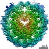 21971MC 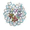 6wz5C  6x0lC  6x0mC 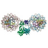 6x0nC C: citing same article ( M: map data used to model this data |
|---|---|
| Similar structure data |


-Links
 PDBj
PDBj


- Assembly
| Deposited unit | 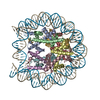
|
|---|---|
| 1 |
|
-Components
-Protein , 4 types, 8 molecules AEBFCGDH
| #1: Protein | Mass: 15303.930 Da / Num. of mol.: 2 Source method: isolated from a genetically manipulated source Source: (gene. exp.)  #2: Protein | Mass: 11263.231 Da / Num. of mol.: 2 Source method: isolated from a genetically manipulated source Source: (gene. exp.) #3: Protein | Mass: 13978.241 Da / Num. of mol.: 2 Source method: isolated from a genetically manipulated source Source: (gene. exp.) #4: Protein | Mass: 13524.752 Da / Num. of mol.: 2 Source method: isolated from a genetically manipulated source Source: (gene. exp.) |
|---|
-DNA chain , 2 types, 2 molecules IJ
| #5: DNA chain | Mass: 51776.004 Da / Num. of mol.: 1 Source method: isolated from a genetically manipulated source Source: (gene. exp.) synthetic construct (others) / Production host: |
|---|---|
| #6: DNA chain | Mass: 51330.684 Da / Num. of mol.: 1 Source method: isolated from a genetically manipulated source Source: (gene. exp.) synthetic construct (others) / Production host: |
-Details
| Has protein modification | N |
|---|
-Experimental details
-Experiment
| Experiment | Method: ELECTRON MICROSCOPY |
|---|---|
| EM experiment | Aggregation state: PARTICLE / 3D reconstruction method: single particle reconstruction |
- Sample preparation
| Component |
| ||||||||||||||||||||||||||||
|---|---|---|---|---|---|---|---|---|---|---|---|---|---|---|---|---|---|---|---|---|---|---|---|---|---|---|---|---|---|
| Molecular weight | Value: 0.24 MDa / Experimental value: NO | ||||||||||||||||||||||||||||
| Source (natural) |
| ||||||||||||||||||||||||||||
| Source (recombinant) |
| ||||||||||||||||||||||||||||
| Buffer solution | pH: 7.5 | ||||||||||||||||||||||||||||
| Specimen | Conc.: 0.1 mg/ml / Embedding applied: NO / Shadowing applied: NO / Staining applied: NO / Vitrification applied: YES | ||||||||||||||||||||||||||||
| Vitrification | Cryogen name: ETHANE |
- Electron microscopy imaging
| Experimental equipment |  Model: Titan Krios / Image courtesy: FEI Company |
|---|---|
| Microscopy | Model: FEI TITAN KRIOS |
| Electron gun | Electron source:  FIELD EMISSION GUN / Accelerating voltage: 300 kV / Illumination mode: FLOOD BEAM FIELD EMISSION GUN / Accelerating voltage: 300 kV / Illumination mode: FLOOD BEAM |
| Electron lens | Mode: BRIGHT FIELD |
| Image recording | Electron dose: 80 e/Å2 / Detector mode: COUNTING / Film or detector model: GATAN K3 BIOQUANTUM (6k x 4k) |
- Processing
| Software | Name: PHENIX / Version: 1.17.1_3660: / Classification: refinement | ||||||||||||||||||||||||
|---|---|---|---|---|---|---|---|---|---|---|---|---|---|---|---|---|---|---|---|---|---|---|---|---|---|
| EM software | Name: PHENIX / Category: model refinement | ||||||||||||||||||||||||
| CTF correction | Type: PHASE FLIPPING AND AMPLITUDE CORRECTION | ||||||||||||||||||||||||
| Symmetry | Point symmetry: C2 (2 fold cyclic) | ||||||||||||||||||||||||
| 3D reconstruction | Resolution: 2.8 Å / Resolution method: FSC 0.143 CUT-OFF / Num. of particles: 934000 / Symmetry type: POINT | ||||||||||||||||||||||||
| Refine LS restraints |
|
