 Movie
Movie Controller

[English] 日本語
 Yorodumi
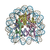
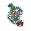
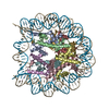
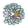
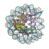
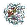
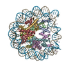
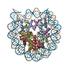
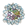
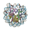
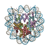
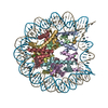
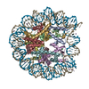
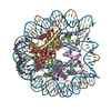
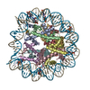
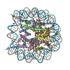
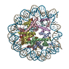
- PDB-4x23: CRYSTAL STRUCTURE OF CENP-C IN COMPLEX WITH THE NUCLEOSOME CORE P... -
+ Open data

- Basic information
| Entry | Database: PDB / ID: 4x23 | |||||||||
|---|---|---|---|---|---|---|---|---|---|---|
| Title | CRYSTAL STRUCTURE OF CENP-C IN COMPLEX WITH THE NUCLEOSOME CORE PARTICLE | |||||||||
 Components Components |
| |||||||||
 Keywords Keywords | structural protein/dna / NUCLEOSOME CORE PARTICLE / WIDOM 601 DNA FRAGMMENT / HISTONE FOLD / CENP-C COMPLEX / SEGREGATION / CHROMOSOME CENTROMERE / KINETOCHORE ASSEMBLY / CONSTITUTIVE CENTROMERE-ASSOCIATED NETWORK (CCAN) PROTEINS / STRUCTURAL PROTEIN-DNA COMPLEX | |||||||||
| Function / homology |  Function and homology information Function and homology informationHDMs demethylate histones / PKMTs methylate histone lysines / Interleukin-7 signaling / Chromatin modifying enzymes / : / SUMOylation of chromatin organization proteins / Metalloprotease DUBs / E3 ubiquitin ligases ubiquitinate target proteins / Factors involved in megakaryocyte development and platelet production / RCAF complex ...HDMs demethylate histones / PKMTs methylate histone lysines / Interleukin-7 signaling / Chromatin modifying enzymes / : / SUMOylation of chromatin organization proteins / Metalloprotease DUBs / E3 ubiquitin ligases ubiquitinate target proteins / Factors involved in megakaryocyte development and platelet production / RCAF complex / RMTs methylate histone arginines / Recruitment and ATM-mediated phosphorylation of repair and signaling proteins at DNA double strand breaks / SIRT1 negatively regulates rRNA expression / NoRC negatively regulates rRNA expression / Activated PKN1 stimulates transcription of AR (androgen receptor) regulated genes KLK2 and KLK3 / polytene chromosome band / PRC2 methylates histones and DNA / HDACs deacetylate histones / Ub-specific processing proteases / Negative Regulation of CDH1 Gene Transcription / Formation of the beta-catenin:TCF transactivating complex / MLL4 and MLL3 complexes regulate expression of PPARG target genes in adipogenesis and hepatic steatosis / RNA Polymerase I Promoter Escape / Regulation of endogenous retroelements by KRAB-ZFP proteins / larval somatic muscle development / RUNX1 regulates genes involved in megakaryocyte differentiation and platelet function / Senescence-Associated Secretory Phenotype (SASP) / Transcriptional regulation by small RNAs / Estrogen-dependent gene expression / HATs acetylate histones / Assembly of the ORC complex at the origin of replication / Oxidative Stress Induced Senescence / UCH proteinases / spindle attachment to meiosis I kinetochore / polytene chromosome / centromeric DNA binding / kinetochore assembly / attachment of mitotic spindle microtubules to kinetochore / condensed chromosome, centromeric region / inner kinetochore / nucleosomal DNA binding / nuclear chromosome / pericentric heterochromatin / chromosome segregation / kinetochore / structural constituent of chromatin / nucleosome / mitotic cell cycle / nucleosome assembly / heterochromatin formation / chromosome / chromatin organization / protein heterodimerization activity / chromatin / protein-containing complex binding / DNA binding / identical protein binding / nucleus Similarity search - Function | |||||||||
| Biological species |  Homo sapiens (human) Homo sapiens (human)  | |||||||||
| Method |  X-RAY DIFFRACTION / SYNCHROTRON / MOLECULAR REPLACEMENT / Resolution: 3.5 Å X-RAY DIFFRACTION / SYNCHROTRON / MOLECULAR REPLACEMENT / Resolution: 3.5 Å | |||||||||
 Authors Authors | Jiang, J.S. | |||||||||
 Citation Citation | Journal: Science / Year: 2013 Title: A conserved mechanism for centromeric nucleosome recognition by centromere protein CENP-C. Authors: Kato, H. / Jiang, J.S. / Zhou, B.R. / Rozendaal, M. / Feng, H. / Ghirlando, R. / Xiao, T.S. / Straight, A.F. / Bai, Y. | |||||||||
| History |
|
- Structure visualization
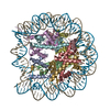
| Structure viewer | Molecule: MolmilJmol/JSmol |
|---|
- Downloads & links
-Download
| PDBx/mmCIF format | 4x23.cif.gz | 618.5 KB | Display | PDBx/mmCIF format |
|---|---|---|---|---|
| PDB format | pdb4x23.ent.gz | 482.9 KB | Display | PDB format |
| PDBx/mmJSON format | 4x23.json.gz | Tree view | PDBx/mmJSON format | |
| Others |  Other downloads Other downloads |
-Validation report
| Arichive directory | https://data.pdbj.org/pub/pdb/validation_reports/x2/4x23ftp://data.pdbj.org/pub/pdb/validation_reports/x2/4x23 | HTTPS FTP |
|---|
-Related structure data


-Links
 PDBj
PDBj


- Assembly
| Deposited unit | 
| ||||||||
|---|---|---|---|---|---|---|---|---|---|
| 1 | 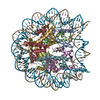
| ||||||||
| 2 | 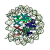
| ||||||||
| Unit cell |
|
-Components
-DNA chain , 2 types, 4 molecules ISJT
| #1: DNA chain | Mass: 45138.770 Da / Num. of mol.: 2 Source method: isolated from a genetically manipulated source Details: 147 BP WIDOM 601 DNA FRAGMENT (+ STRAND) / Source: (gene. exp.) Homo sapiens (human) / Production host:  #2: DNA chain | Mass: 45610.043 Da / Num. of mol.: 2 Source method: isolated from a genetically manipulated source Details: 147 BP WIDOM 601 DNA FRAGMENT (- STRAND) / Source: (gene. exp.) Homo sapiens (human) / Production host: |
|---|
-Protein , 4 types, 16 molecules AEKOBFLPCGMQDHNR
| #3: Protein | Mass: 11365.296 Da / Num. of mol.: 4 / Fragment: UNP RESIDUES 41-133 Source method: isolated from a genetically manipulated source Source: (gene. exp.) #4: Protein | Mass: 8910.394 Da / Num. of mol.: 4 / Fragment: UNP RESIDUES 25-103 Source method: isolated from a genetically manipulated source Source: (gene. exp.) #5: Protein | Mass: 11093.877 Da / Num. of mol.: 4 / Fragment: UNP RESIDUES 16-117 Source method: isolated from a genetically manipulated source Source: (gene. exp.) #6: Protein | Mass: 10019.455 Da / Num. of mol.: 4 / Fragment: UNP RESIDUES 33-122 Source method: isolated from a genetically manipulated source Source: (gene. exp.) |
|---|
-Protein/peptide , 1 types, 4 molecules VUXW
| #7: Protein/peptide | Mass: 3222.686 Da / Num. of mol.: 4 / Fragment: UNP RESIDUES 710-734 / Source method: obtained synthetically / Source: (synth.) |
|---|
-Details
| Sequence details | THE DISCREPANCY AT THE C-TERM OF H3 SEQUENCES (CHAINS A,E,K,O) IS A RESULT OF CHIMERIC CENP-A, I.E. ...THE DISCREPANC |
|---|
-Experimental details
-Experiment
| Experiment | Method: X-RAY DIFFRACTION |
|---|
- Sample preparation
| Crystal | Density Matthews: 2.63 Å3/Da / Density % sol: 53.25 % |
|---|---|
| Crystal grow | Temperature: 294 K / Method: vapor diffusion, hanging drop / pH: 7.5 Details: 10% MPD, 40MM SODIUM CACODYLATE, 24MM SPERMINE TETRA-HCL, 80MM SODIUM CHLORIDE, 20MM MAGNESIUM CHLORIDE; RESERVIOR 35% MPD. PH 7.5, VAPOR DIFFUSION, HANGING DROP, TEMPERATURE 294K PH range: 7.4 - 7.6 |
-Data collection
| Diffraction |
| ||||||||||||||||||
|---|---|---|---|---|---|---|---|---|---|---|---|---|---|---|---|---|---|---|---|
| Diffraction source |
| ||||||||||||||||||
| Detector |
| ||||||||||||||||||
| Radiation |
| ||||||||||||||||||
| Radiation wavelength |
| ||||||||||||||||||
| Reflection | Resolution: 3.5→50 Å / Num. obs: 48623 / % possible obs: 96.1 % / Observed criterion σ(F): 0 / Observed criterion σ(I): -3 / Redundancy: 4 % / Biso Wilson estimate: 122.9 Å2 / Rmerge(I) obs: 0.118 / Net I/σ(I): 11 | ||||||||||||||||||
| Reflection shell | Resolution: 3.5→3.56 Å / Redundancy: 3 % / Rmerge(I) obs: 0.695 / Mean I/σ(I) obs: 1.5 / % possible all: 78.7 |

- Processing
| Software |
| ||||||||||||||||||||||||||||||||||||||||||||||||||||||||||||||||||||||||||||||||||||||||||||||||||
|---|---|---|---|---|---|---|---|---|---|---|---|---|---|---|---|---|---|---|---|---|---|---|---|---|---|---|---|---|---|---|---|---|---|---|---|---|---|---|---|---|---|---|---|---|---|---|---|---|---|---|---|---|---|---|---|---|---|---|---|---|---|---|---|---|---|---|---|---|---|---|---|---|---|---|---|---|---|---|---|---|---|---|---|---|---|---|---|---|---|---|---|---|---|---|---|---|---|---|---|
| Refinement | Method to determine structure: MOLECULAR REPLACEMENT Starting model: 2PYO, 3MVD Resolution: 3.5→49.543 Å / SU ML: 0.59 / Cross valid method: THROUGHOUT / σ(F): 1.33 / Phase error: 33.82 / Stereochemistry target values: ML / Details: CNS 1.3 WAS USED FOR LOW RESOLUTION REFINEMENT
| ||||||||||||||||||||||||||||||||||||||||||||||||||||||||||||||||||||||||||||||||||||||||||||||||||
| Solvent computation | Shrinkage radii: 0.9 Å / VDW probe radii: 1.11 Å / Solvent model: FLAT BULK SOLVENT MODEL | ||||||||||||||||||||||||||||||||||||||||||||||||||||||||||||||||||||||||||||||||||||||||||||||||||
| Displacement parameters | Biso mean: 157.6 Å2 | ||||||||||||||||||||||||||||||||||||||||||||||||||||||||||||||||||||||||||||||||||||||||||||||||||
| Refinement step | Cycle: LAST / Resolution: 3.5→49.543 Å
| ||||||||||||||||||||||||||||||||||||||||||||||||||||||||||||||||||||||||||||||||||||||||||||||||||
| Refine LS restraints |
| ||||||||||||||||||||||||||||||||||||||||||||||||||||||||||||||||||||||||||||||||||||||||||||||||||
| LS refinement shell |
|
