 Movie
Movie Controller

[English] 日本語
 Yorodumi
- PDB-1u35: Crystal structure of the nucleosome core particle containing the ... -
+ Open data

- Basic information
| Entry | Database: PDB / ID: 1u35 | ||||||
|---|---|---|---|---|---|---|---|
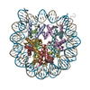
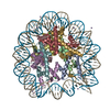
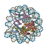
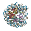
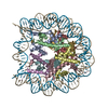
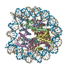
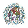
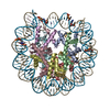
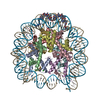
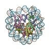
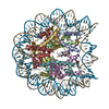
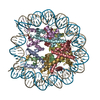
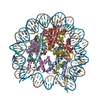
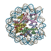
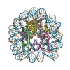
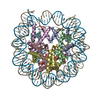
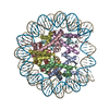
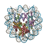
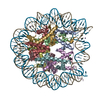
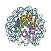
| Title | Crystal structure of the nucleosome core particle containing the histone domain of macroH2A | ||||||
 Components Components |
| ||||||
 Keywords Keywords | structural protein/DNA / Nucleosome / NCP / Histone fold / Histone variant / macroH2A / structural protein-DNA COMPLEX | ||||||
| Function / homology |  Function and homology information Function and homology informationRegulation of PD-L1(CD274) transcription / negative regulation of cell cycle G2/M phase transition / negative regulation of protein localization to chromosome, telomeric region / regulation of NAD metabolic process / positive regulation of response to oxidative stress / Deposition of new CENPA-containing nucleosomes at the centromere / Inhibition of DNA recombination at telomere / positive regulation of maintenance of mitotic sister chromatid cohesion / SUMOylation of chromatin organization proteins / DNA Damage/Telomere Stress Induced Senescence ...Regulation of PD-L1(CD274) transcription / negative regulation of cell cycle G2/M phase transition / negative regulation of protein localization to chromosome, telomeric region / regulation of NAD metabolic process / positive regulation of response to oxidative stress / Deposition of new CENPA-containing nucleosomes at the centromere / Inhibition of DNA recombination at telomere / positive regulation of maintenance of mitotic sister chromatid cohesion / SUMOylation of chromatin organization proteins / DNA Damage/Telomere Stress Induced Senescence / Chromatin modifying enzymes / G2/M DNA damage checkpoint / Interleukin-7 signaling / regulation of response to oxidative stress / Regulation of endogenous retroelements by KRAB-ZFP proteins / ADP-D-ribose binding / ADP-D-ribose modification-dependent protein binding / Condensation of Prophase Chromosomes / HDMs demethylate histones / Nonhomologous End-Joining (NHEJ) / Negative Regulation of CDH1 Gene Transcription / negative regulation of transcription of nucleolar large rRNA by RNA polymerase I / HDACs deacetylate histones / Recognition and association of DNA glycosylase with site containing an affected purine / Cleavage of the damaged purine / PRC2 methylates histones and DNA / HATs acetylate histones / MLL4 and MLL3 complexes regulate expression of PPARG target genes in adipogenesis and hepatic steatosis / PKMTs methylate histone lysines / Processing of DNA double-strand break ends / Recruitment and ATM-mediated phosphorylation of repair and signaling proteins at DNA double strand breaks / RUNX1 regulates genes involved in megakaryocyte differentiation and platelet function / double-stranded methylated DNA binding / positive regulation of endodermal cell differentiation / regulation of oxidative phosphorylation / establishment of protein localization to chromatin / RMTs methylate histone arginines / Factors involved in megakaryocyte development and platelet production / Barr body / sex chromatin / Estrogen-dependent gene expression / rDNA binding / dosage compensation by inactivation of X chromosome / poly-ADP-D-ribose modification-dependent protein binding / positive regulation of keratinocyte differentiation / negative regulation of response to oxidative stress / nucleosomal DNA binding / protein poly-ADP-ribosylation / nuclear chromosome / negative regulation of gene expression, epigenetic / regulation of lipid metabolic process / protein serine/threonine kinase inhibitor activity / pericentric heterochromatin / site of DNA damage / protein localization to CENP-A containing chromatin / condensed chromosome / CENP-A containing nucleosome / epigenetic regulation of gene expression / transcription initiation-coupled chromatin remodeling / RNA polymerase II transcription regulatory region sequence-specific DNA binding / promoter-specific chromatin binding / chromatin DNA binding / structural constituent of chromatin / nucleosome / nucleosome assembly / heterochromatin formation / chromatin organization / chromosome, telomeric region / transcription cis-regulatory region binding / protein heterodimerization activity / DNA repair / protein kinase binding / chromatin / nucleolus / enzyme binding / negative regulation of transcription by RNA polymerase II / protein-containing complex / DNA binding / extracellular exosome / nucleoplasm / nucleus Similarity search - Function | ||||||
| Biological species |  Homo sapiens (human) Homo sapiens (human) | ||||||
| Method |  X-RAY DIFFRACTION / SYNCHROTRON / MOLECULAR REPLACEMENT / Resolution: 3 Å X-RAY DIFFRACTION / SYNCHROTRON / MOLECULAR REPLACEMENT / Resolution: 3 Å | ||||||
 Authors Authors | Chakravarthy, S. / Gundimella, S.K. / Caron, C. / Perche, P.Y. / Pehrson, J.R. / Khochbin, S. / Luger, K. | ||||||
 Citation Citation | Journal: Mol.Cell.Biol. / Year: 2005 Title: Structural characterization of the histone variant macroH2A. Authors: Chakravarthy, S. / Gundimella, S.K. / Caron, C. / Perche, P.Y. / Pehrson, J.R. / Khochbin, S. / Luger, K. | ||||||
| History |
|
- Structure visualization
| Structure viewer | Molecule: MolmilJmol/JSmol |
|---|
- Downloads & links
-Download
| PDBx/mmCIF format | 1u35.cif.gz | 318.1 KB | Display | PDBx/mmCIF format |
|---|---|---|---|---|
| PDB format | pdb1u35.ent.gz | 243.4 KB | Display | PDB format |
| PDBx/mmJSON format | 1u35.json.gz | Tree view | PDBx/mmJSON format | |
| Others |  Other downloads Other downloads |
-Validation report
| Arichive directory | https://data.pdbj.org/pub/pdb/validation_reports/u3/1u35ftp://data.pdbj.org/pub/pdb/validation_reports/u3/1u35 | HTTPS FTP |
|---|
-Related structure data
| Related structure data | 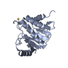 1yd9C 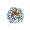 1aoiS S: Starting model for refinement C: citing same article ( |
|---|---|
| Similar structure data |

-Links
 PDBj
PDBj


- Assembly
| Deposited unit | 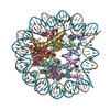
| ||||||||
|---|---|---|---|---|---|---|---|---|---|
| 1 |
| ||||||||
| Unit cell |
|
-Components
-Protein , 4 types, 8 molecules AEBFCGDH
| #2: Protein | Mass: 15437.167 Da / Num. of mol.: 2 Source method: isolated from a genetically manipulated source Source: (gene. exp.)  #3: Protein | Mass: 11394.426 Da / Num. of mol.: 2 Source method: isolated from a genetically manipulated source Source: (gene. exp.) #4: Protein | Mass: 12984.343 Da / Num. of mol.: 2 Source method: isolated from a genetically manipulated source Source: (gene. exp.) Homo sapiens (human) / Plasmid: pet3a / Production host: #5: Protein | Mass: 14025.280 Da / Num. of mol.: 2 Source method: isolated from a genetically manipulated source Source: (gene. exp.) |
|---|
-DNA chain / Non-polymers , 2 types, 107 molecules IJ
| #1: DNA chain | Mass: 45054.844 Da / Num. of mol.: 2 Source method: isolated from a genetically manipulated source Source: (gene. exp.) Homo sapiens (human) / Plasmid: puc19 / Production host: #6: Water | ChemComp-HOH / | Mass: 18.015 Da / Num. of mol.: 105 / Source method: isolated from a natural source / Formula: H2O |
|---|
-Experimental details
-Experiment
| Experiment | Method: X-RAY DIFFRACTION / Number of used crystals: 1 |
|---|
- Sample preparation
| Crystal | Density Matthews: 2.5 Å3/Da / Density % sol: 52 % | ||||||||||||||||||||||||||||||||||||
|---|---|---|---|---|---|---|---|---|---|---|---|---|---|---|---|---|---|---|---|---|---|---|---|---|---|---|---|---|---|---|---|---|---|---|---|---|---|
| Crystal grow | Temperature: 292 K / Method: vapor diffusion, sitting drop / pH: 6 Details: potassium chloride, manganese chloride, potassium cacodylate, pH 6.0, VAPOR DIFFUSION, SITTING DROP, temperature 292K | ||||||||||||||||||||||||||||||||||||
| Components of the solutions |
|
-Data collection
| Diffraction | Mean temperature: 298 K |
|---|---|
| Diffraction source | Source: SYNCHROTRON / Site: ALS  / Beamline: 8.2.2 / Wavelength: 1.1 Å / Beamline: 8.2.2 / Wavelength: 1.1 Å |
| Detector | Type: ADSC QUANTUM 210 / Detector: CCD / Date: Feb 27, 2003 |
| Radiation | Protocol: SINGLE WAVELENGTH / Monochromatic (M) / Laue (L): M / Scattering type: x-ray |
| Radiation wavelength | Wavelength: 1.1 Å / Relative weight: 1 |
| Reflection | Resolution: 2.95→50 Å / Num. obs: 43366 / % possible obs: 99.5 % / Observed criterion σ(I): 2 / Rmerge(I) obs: 0.095 |
| Reflection shell | Resolution: 2.95→3.02 Å / Rmerge(I) obs: 0.421 / Mean I/σ(I) obs: 3.15 / Num. unique all: 2865 / % possible all: 100 |
- Processing
| Software |
| |||||||||||||||||||||||||
|---|---|---|---|---|---|---|---|---|---|---|---|---|---|---|---|---|---|---|---|---|---|---|---|---|---|---|
| Refinement | Method to determine structure: MOLECULAR REPLACEMENT Starting model: pdb entry 1AOI Resolution: 3→50 Å / σ(F): 0 / Stereochemistry target values: Engh & Huber Details: There are close contacts between A217 and T218 in chain J, between T74 and C75 in chain I.
| |||||||||||||||||||||||||
| Refinement step | Cycle: LAST / Resolution: 3→50 Å
| |||||||||||||||||||||||||
| Refine LS restraints |
|
