 Movie
Movie Controller

[English] 日本語
 Yorodumi
- PDB-1aoi: COMPLEX BETWEEN NUCLEOSOME CORE PARTICLE (H3,H4,H2A,H2B) AND 146 ... -
+ Open data

- Basic information
| Entry | Database: PDB / ID: 1aoi | ||||||
|---|---|---|---|---|---|---|---|
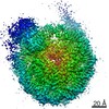
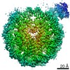
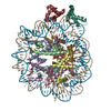
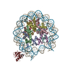
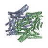
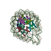
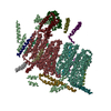
| Title | COMPLEX BETWEEN NUCLEOSOME CORE PARTICLE (H3,H4,H2A,H2B) AND 146 BP LONG DNA FRAGMENT | ||||||
 Components Components |
| ||||||
 Keywords Keywords | DNA BINDING PROTEIN/DNA / NUCLEOSOME / CHROMATIN / HISTONE / PROTEIN DNA INTERACTION / NUCLEOPROTEIN / SUPERCOILED DNA / DNA BINDING PROTEIN-DNA COMPLEX | ||||||
| Function / homology |  Function and homology information Function and homology informationstructural constituent of chromatin / nucleosome / nucleosome assembly / heterochromatin formation / protein heterodimerization activity / DNA binding / nucleus Similarity search - Function | ||||||
| Biological species | |||||||
| Method |  X-RAY DIFFRACTION / SYNCHROTRON / MIR / Resolution: 2.8 Å X-RAY DIFFRACTION / SYNCHROTRON / MIR / Resolution: 2.8 Å | ||||||
 Authors Authors | Luger, K. / Maeder, A.W. / Richmond, R.K. / Sargent, D.F. / Richmond, T.J. | ||||||
 Citation Citation | Journal: Nature / Year: 1997 Title: Crystal structure of the nucleosome core particle at 2.8 A resolution. Authors: Luger, K. / Mader, A.W. / Richmond, R.K. / Sargent, D.F. / Richmond, T.J. | ||||||
| History |
|
- Structure visualization
| Structure viewer | Molecule: MolmilJmol/JSmol |
|---|
- Downloads & links
-Download
| PDBx/mmCIF format | 1aoi.cif.gz | 294 KB | Display | PDBx/mmCIF format |
|---|---|---|---|---|
| PDB format | pdb1aoi.ent.gz | 228.4 KB | Display | PDB format |
| PDBx/mmJSON format | 1aoi.json.gz | Tree view | PDBx/mmJSON format | |
| Others |  Other downloads Other downloads |
-Validation report
| Arichive directory | https://data.pdbj.org/pub/pdb/validation_reports/ao/1aoiftp://data.pdbj.org/pub/pdb/validation_reports/ao/1aoi | HTTPS FTP |
|---|
-Related structure data
| Similar structure data |
|---|

-Links
 PDBj
PDBj


- Assembly
| Deposited unit | 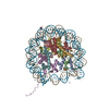
| ||||||||||
|---|---|---|---|---|---|---|---|---|---|---|---|
| 1 |
| ||||||||||
| Unit cell |
|
-Components
-DNA chain , 1 types, 2 molecules IJ
| #1: DNA chain | Mass: 45053.855 Da / Num. of mol.: 2 / Source method: obtained synthetically |
|---|
-Protein , 4 types, 8 molecules AEBFCGDH
| #2: Protein | Mass: 13244.528 Da / Num. of mol.: 2 / Fragment: HISTONE H3 Source method: isolated from a genetically manipulated source Source: (gene. exp.)  #3: Protein | Mass: 9990.770 Da / Num. of mol.: 2 / Fragment: HISTONE H4 Source method: isolated from a genetically manipulated source Source: (gene. exp.) Description: SYNTHETIC GENE, OPTIMIZED CODON USAGE FOR ESCHERICHIA COLI; Production host: #4: Protein | Mass: 12639.772 Da / Num. of mol.: 2 / Fragment: HISTONE H2A Source method: isolated from a genetically manipulated source Source: (gene. exp.) #5: Protein | Mass: 11179.959 Da / Num. of mol.: 2 / Fragment: HISTONE H2B / Mutation: A7P Source method: isolated from a genetically manipulated source Source: (gene. exp.) |
|---|
-Non-polymers , 2 types, 19 molecules 

| #6: Chemical | ChemComp-MN /  Mass: 54.938 Da / Num. of mol.: 6 / Source method: obtained synthetically / Formula: Mn Mass: 54.938 Da / Num. of mol.: 6 / Source method: obtained synthetically / Formula: Mn#7: Water | ChemComp-HOH / | Mass: 18.015 Da / Num. of mol.: 13 / Source method: isolated from a natural source / Formula: H2O |
|---|
-Experimental details
-Experiment
| Experiment | Method: X-RAY DIFFRACTION / Number of used crystals: 1 |
|---|
- Sample preparation
| Crystal | Density Matthews: 2.83 Å3/Da / Density % sol: 56.54 % | ||||||||||||||||||||||||||||||||||||||||||||||||||||||||
|---|---|---|---|---|---|---|---|---|---|---|---|---|---|---|---|---|---|---|---|---|---|---|---|---|---|---|---|---|---|---|---|---|---|---|---|---|---|---|---|---|---|---|---|---|---|---|---|---|---|---|---|---|---|---|---|---|---|
| Crystal grow | *PLUS Temperature: 20 ℃ / pH: 6 / Method: vapor diffusion | ||||||||||||||||||||||||||||||||||||||||||||||||||||||||
| Components of the solutions | *PLUS
|
-Data collection
| Diffraction | Mean temperature: 110 K |
|---|---|
| Diffraction source | Source: SYNCHROTRON / Site: ESRF  / Beamline: ID13 / Beamline: ID13 |
| Detector | Type: MARRESEARCH / Detector: IMAGE PLATE / Date: Aug 15, 1995 |
| Radiation | Protocol: SINGLE WAVELENGTH / Monochromatic (M) / Laue (L): M / Scattering type: x-ray |
| Radiation wavelength | Relative weight: 1 |
| Reflection | Resolution: 2.8→25 Å / Num. obs: 52487 / % possible obs: 99 % / Redundancy: 5.7 % / Rmerge(I) obs: 0.056 / Rsym value: 3.6 |
| Reflection shell | Resolution: 2.8→2.85 Å / Redundancy: 6.3 % / Rmerge(I) obs: 0.132 / % possible all: 95 |
| Reflection | *PLUS Num. measured all: 297397 |
- Processing
| Software |
| ||||||||||||||||||||||||||||||||||||||||||||||||||||||||||||
|---|---|---|---|---|---|---|---|---|---|---|---|---|---|---|---|---|---|---|---|---|---|---|---|---|---|---|---|---|---|---|---|---|---|---|---|---|---|---|---|---|---|---|---|---|---|---|---|---|---|---|---|---|---|---|---|---|---|---|---|---|---|
| Refinement | Method to determine structure: MIR / Resolution: 2.8→25 Å / Isotropic thermal model: RESTRAINED / Cross valid method: THROUGHOUT / Details: BULK SOLVENT MODEL USED
| ||||||||||||||||||||||||||||||||||||||||||||||||||||||||||||
| Solvent computation | Solvent model: BULK SOLVENT MODEL USED | ||||||||||||||||||||||||||||||||||||||||||||||||||||||||||||
| Refinement step | Cycle: LAST / Resolution: 2.8→25 Å
| ||||||||||||||||||||||||||||||||||||||||||||||||||||||||||||
| Refine LS restraints |
| ||||||||||||||||||||||||||||||||||||||||||||||||||||||||||||
| Software | *PLUS Name: X-PLOR / Version: 3.843 / Classification: refinement | ||||||||||||||||||||||||||||||||||||||||||||||||||||||||||||
| Refinement | *PLUS σ(F): 3 / Rfactor obs: 0.224 | ||||||||||||||||||||||||||||||||||||||||||||||||||||||||||||
| Solvent computation | *PLUS | ||||||||||||||||||||||||||||||||||||||||||||||||||||||||||||
| Displacement parameters | *PLUS |
