 Movie
Movie Controller


+ Open data

- Basic information
| Entry | Database: PDB / ID: 2f8n | ||||||
|---|---|---|---|---|---|---|---|
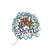
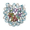
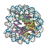
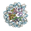
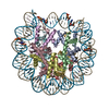
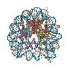
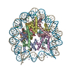
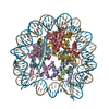
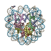
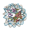
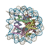
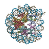
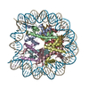
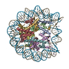
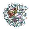
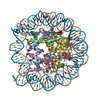
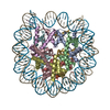
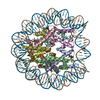
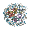
| Title | 2.9 Angstrom X-ray structure of hybrid macroH2A nucleosomes | ||||||
 Components Components |
| ||||||
 Keywords Keywords | STRUCTURAL PROTEIN/DNA / Nucleosome / NCP / macroH2A / Histone variant / chromatin / STRUCTURAL PROTEIN-DNA COMPLEX | ||||||
| Function / homology |  Function and homology information Function and homology informationRegulation of PD-L1(CD274) transcription / negative regulation of cell cycle G2/M phase transition / negative regulation of protein localization to chromosome, telomeric region / regulation of NAD metabolic process / positive regulation of response to oxidative stress / Deposition of new CENPA-containing nucleosomes at the centromere / Inhibition of DNA recombination at telomere / positive regulation of maintenance of mitotic sister chromatid cohesion / DNA Damage/Telomere Stress Induced Senescence / regulation of response to oxidative stress ...Regulation of PD-L1(CD274) transcription / negative regulation of cell cycle G2/M phase transition / negative regulation of protein localization to chromosome, telomeric region / regulation of NAD metabolic process / positive regulation of response to oxidative stress / Deposition of new CENPA-containing nucleosomes at the centromere / Inhibition of DNA recombination at telomere / positive regulation of maintenance of mitotic sister chromatid cohesion / DNA Damage/Telomere Stress Induced Senescence / regulation of response to oxidative stress / Regulation of endogenous retroelements by KRAB-ZFP proteins / ADP-D-ribose binding / ADP-D-ribose modification-dependent protein binding / Condensation of Prophase Chromosomes / Negative Regulation of CDH1 Gene Transcription / negative regulation of transcription of nucleolar large rRNA by RNA polymerase I / Recognition and association of DNA glycosylase with site containing an affected purine / Cleavage of the damaged purine / PRC2 methylates histones and DNA / Metalloprotease DUBs / MLL4 and MLL3 complexes regulate expression of PPARG target genes in adipogenesis and hepatic steatosis / UCH proteinases / RUNX1 regulates genes involved in megakaryocyte differentiation and platelet function / double-stranded methylated DNA binding / positive regulation of endodermal cell differentiation / regulation of oxidative phosphorylation / establishment of protein localization to chromatin / RMTs methylate histone arginines / Barr body / sex chromatin / rDNA binding / dosage compensation by inactivation of X chromosome / poly-ADP-D-ribose modification-dependent protein binding / Ub-specific processing proteases / positive regulation of keratinocyte differentiation / negative regulation of response to oxidative stress / nucleosomal DNA binding / protein poly-ADP-ribosylation / nuclear chromosome / negative regulation of gene expression, epigenetic / regulation of lipid metabolic process / protein serine/threonine kinase inhibitor activity / pericentric heterochromatin / site of DNA damage / condensed chromosome / epigenetic regulation of gene expression / transcription initiation-coupled chromatin remodeling / RNA polymerase II transcription regulatory region sequence-specific DNA binding / promoter-specific chromatin binding / chromatin DNA binding / structural constituent of chromatin / nucleosome / nucleosome assembly / heterochromatin formation / chromosome, telomeric region / transcription cis-regulatory region binding / protein heterodimerization activity / DNA repair / protein kinase binding / chromatin / nucleolus / enzyme binding / negative regulation of transcription by RNA polymerase II / DNA binding / extracellular exosome / nucleoplasm / nucleus Similarity search - Function | ||||||
| Biological species |  Homo sapiens (human) Homo sapiens (human) | ||||||
| Method |  X-RAY DIFFRACTION / SYNCHROTRON / MOLECULAR REPLACEMENT / Resolution: 2.9 Å X-RAY DIFFRACTION / SYNCHROTRON / MOLECULAR REPLACEMENT / Resolution: 2.9 Å | ||||||
 Authors Authors | Chakravarthy, S. / Luger, K. | ||||||
 Citation Citation | Journal: To be Published Title: Nucleosomes containing the histone domain of macroH2A: In vitro possibilities. Authors: Chakravarthy, S. / Luger, K. | ||||||
| History |
|
- Structure visualization
| Structure viewer | Molecule: MolmilJmol/JSmol |
|---|
- Downloads & links
-Download
| PDBx/mmCIF format | 2f8n.cif.gz | 320.8 KB | Display | PDBx/mmCIF format |
|---|---|---|---|---|
| PDB format | pdb2f8n.ent.gz | 244.8 KB | Display | PDB format |
| PDBx/mmJSON format | 2f8n.json.gz | Tree view | PDBx/mmJSON format | |
| Others |  Other downloads Other downloads |
-Validation report
| Arichive directory | https://data.pdbj.org/pub/pdb/validation_reports/f8/2f8nftp://data.pdbj.org/pub/pdb/validation_reports/f8/2f8n | HTTPS FTP |
|---|
-Related structure data
| Related structure data |  1u35S S: Starting model for refinement |
|---|---|
| Similar structure data |


-Links
 PDBj
PDBj


- Assembly
| Deposited unit | 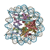
| ||||||||
|---|---|---|---|---|---|---|---|---|---|
| 1 |
| ||||||||
| Unit cell |
| ||||||||
| Details | The biological assembly is an octamer of histones wrapped by 146 basepairs of DNA called the nucleosome core particle, which is also the asymmetric unit. (all of which, the coordinates are given for in the submitted pdb file). |
-Components
-Protein , 6 types, 8 molecules AEBFDHGK
| #2: Protein | Mass: 15421.101 Da / Num. of mol.: 2 Source method: isolated from a genetically manipulated source Source: (gene. exp.)  #3: Protein | Mass: 11394.426 Da / Num. of mol.: 2 Source method: isolated from a genetically manipulated source Source: (gene. exp.) #4: Protein | | Mass: 14025.280 Da / Num. of mol.: 1 Source method: isolated from a genetically manipulated source Source: (gene. exp.) #5: Protein | | Mass: 13655.948 Da / Num. of mol.: 1 Source method: isolated from a genetically manipulated source Source: (gene. exp.) #6: Protein | | Mass: 12984.343 Da / Num. of mol.: 1 / Fragment: residues 0-119 Source method: isolated from a genetically manipulated source Source: (gene. exp.) Homo sapiens (human) / Plasmid: pet3a / Production host: #7: Protein | | Mass: 16198.775 Da / Num. of mol.: 1 Source method: isolated from a genetically manipulated source Source: (gene. exp.) |
|---|
-DNA chain / Non-polymers , 2 types, 122 molecules IJ
| #1: DNA chain | Mass: 45054.844 Da / Num. of mol.: 2 Source method: isolated from a genetically manipulated source Source: (gene. exp.) Homo sapiens (human) / Plasmid: puc19 / Production host: #8: Water | ChemComp-HOH / | Mass: 18.015 Da / Num. of mol.: 120 / Source method: isolated from a natural source / Formula: H2O |
|---|
-Experimental details
-Experiment
| Experiment | Method: X-RAY DIFFRACTION / Number of used crystals: 1 |
|---|
- Sample preparation
| Crystal | Density Matthews: 2.51 Å3/Da / Density % sol: 50.92 % | ||||||||||||||||||||||||||||||||||||
|---|---|---|---|---|---|---|---|---|---|---|---|---|---|---|---|---|---|---|---|---|---|---|---|---|---|---|---|---|---|---|---|---|---|---|---|---|---|
| Crystal grow | Temperature: 292 K / Method: vapor diffusion, sitting drop / pH: 6 Details: 34 to 37.5mM KCl and 40-45mM MnCl2, 5mM Potassium Cacodylate, Sample concentration: 8-12 mg/ml, pH 6.0, VAPOR DIFFUSION, SITTING DROP, temperature 292K | ||||||||||||||||||||||||||||||||||||
| Components of the solutions |
|
-Data collection
| Diffraction | Mean temperature: 93 K |
|---|---|
| Diffraction source | Source: SYNCHROTRON / Site: ALS  / Beamline: 5.0.2 / Wavelength: 1 Å / Beamline: 5.0.2 / Wavelength: 1 Å |
| Detector | Type: ADSC QUANTUM 210 / Detector: CCD / Date: Feb 10, 2004 |
| Radiation | Protocol: SINGLE WAVELENGTH / Monochromatic (M) / Laue (L): M / Scattering type: x-ray |
| Radiation wavelength | Wavelength: 1 Å / Relative weight: 1 |
| Reflection | Resolution: 2.9→31.4 Å / Num. obs: 44768 / Observed criterion σ(I): 2 / Rmerge(I) obs: 0.07 |
| Reflection shell | Highest resolution: 2.9 Å / Rmerge(I) obs: 0.408 |
- Processing
| Software |
| ||||||||||||||||||||
|---|---|---|---|---|---|---|---|---|---|---|---|---|---|---|---|---|---|---|---|---|---|
| Refinement | Method to determine structure: MOLECULAR REPLACEMENT Starting model: pdb entry 1U35 Resolution: 2.9→31.4 Å / Stereochemistry target values: Engh & Huber Details: A 73 chain I and T 74 chain I are linked together. A 217 chain J and T 218 chain J are linked together. However there are T 73A chain I and A 217A chain J present in the structure. The ...Details: A 73 chain I and T 74 chain I are linked together. A 217 chain J and T 218 chain J are linked together. However there are T 73A chain I and A 217A chain J present in the structure. The electron density for this base pair is lost as a result of a convolution between two stretch conformations on the two halves of the nucleosome on either side of the diad axis.
| ||||||||||||||||||||
| Refinement step | Cycle: LAST / Resolution: 2.9→31.4 Å
| ||||||||||||||||||||
| Refine LS restraints |
|
