 Movie
Movie Controller

[English] 日本語
 Yorodumi
- PDB-2fj7: Crystal structure of Nucleosome Core Particle Containing a Poly (... -
+ Open data

- Basic information
| Entry | Database: PDB / ID: 2fj7 | ||||||
|---|---|---|---|---|---|---|---|
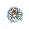
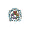
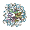
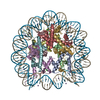
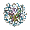
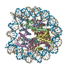
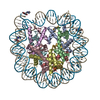
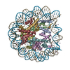
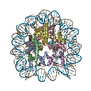
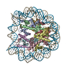
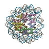
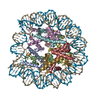
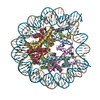
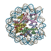
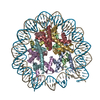
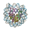
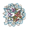
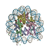
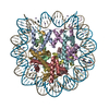
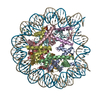
| Title | Crystal structure of Nucleosome Core Particle Containing a Poly (dA.dT) Sequence Element | ||||||
 Components Components |
| ||||||
 Keywords Keywords | STRUCTURAL PROTEIN/DNA / Protein-DNA complex / narrow minor groove / STRUCTURAL PROTEIN-DNA COMPLEX | ||||||
| Function / homology |  Function and homology information Function and homology informationstructural constituent of chromatin / nucleosome / nucleosome assembly / heterochromatin formation / protein heterodimerization activity / DNA binding / nucleoplasm / nucleus Similarity search - Function | ||||||
| Biological species | |||||||
| Method |  X-RAY DIFFRACTION / SYNCHROTRON / MOLECULAR REPLACEMENT / Resolution: 3.2 Å X-RAY DIFFRACTION / SYNCHROTRON / MOLECULAR REPLACEMENT / Resolution: 3.2 Å | ||||||
 Authors Authors | Bao, Y. / White, C.L. / Luger, K. | ||||||
 Citation Citation | Journal: J.Mol.Biol. / Year: 2006 Title: Nucleosome Core Particles Containing a Poly(dA.dT) Sequence Element Exhibit a Locally Distorted DNA Structure. Authors: Bao, Y. / White, C.L. / Luger, K. | ||||||
| History |
|
- Structure visualization
| Structure viewer | Molecule: MolmilJmol/JSmol |
|---|
- Downloads & links
-Download
| PDBx/mmCIF format | 2fj7.cif.gz | 290.8 KB | Display | PDBx/mmCIF format |
|---|---|---|---|---|
| PDB format | pdb2fj7.ent.gz | 222.6 KB | Display | PDB format |
| PDBx/mmJSON format | 2fj7.json.gz | Tree view | PDBx/mmJSON format | |
| Others |  Other downloads Other downloads |
-Validation report
| Arichive directory | https://data.pdbj.org/pub/pdb/validation_reports/fj/2fj7ftp://data.pdbj.org/pub/pdb/validation_reports/fj/2fj7 | HTTPS FTP |
|---|
-Related structure data
| Related structure data | |
|---|---|
| Similar structure data |


-Links
 PDBj
PDBj


- Assembly
| Deposited unit | 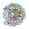
| ||||||||
|---|---|---|---|---|---|---|---|---|---|
| 1 |
| ||||||||
| Unit cell |
| ||||||||
| Details | Histone octomer and the 147 bp DNA containing Poly (dA.dT) element were reconstituted to form NCP, which is the biological unit. |
-Components
-147 bp DNA containing 16 bp poly ... , 2 types, 2 molecules IJ
| #1: DNA chain | Mass: 45369.137 Da / Num. of mol.: 1 / Source method: obtained synthetically |
|---|---|
| #2: DNA chain | Mass: 45350.035 Da / Num. of mol.: 1 / Source method: obtained synthetically |
-Protein , 4 types, 8 molecules AEBFCGDH
| #3: Protein | Mass: 15303.930 Da / Num. of mol.: 2 Source method: isolated from a genetically manipulated source Source: (gene. exp.)  #4: Protein | Mass: 11263.231 Da / Num. of mol.: 2 Source method: isolated from a genetically manipulated source Source: (gene. exp.) #5: Protein | Mass: 13978.241 Da / Num. of mol.: 2 Source method: isolated from a genetically manipulated source Source: (gene. exp.) #6: Protein | Mass: 13848.097 Da / Num. of mol.: 2 Source method: isolated from a genetically manipulated source Source: (gene. exp.) |
|---|
-Experimental details
-Experiment
| Experiment | Method: X-RAY DIFFRACTION / Number of used crystals: 1 |
|---|
- Sample preparation
| Crystal | Density Matthews: 2.52 Å3/Da / Density % sol: 51.26 % | ||||||||||||||||||||||||||||||||||||
|---|---|---|---|---|---|---|---|---|---|---|---|---|---|---|---|---|---|---|---|---|---|---|---|---|---|---|---|---|---|---|---|---|---|---|---|---|---|
| Crystal grow | Temperature: 292 K / Method: vapor diffusion, sitting drop / pH: 6 Details: 20 to 35 mM KCl, 34 to 48 mM MnCl2, and 5mM K-cacodylate pH 6.0 , VAPOR DIFFUSION, SITTING DROP, temperature 292K | ||||||||||||||||||||||||||||||||||||
| Components of the solutions |
|
-Data collection
| Diffraction | Mean temperature: 103 K |
|---|---|
| Diffraction source | Source: SYNCHROTRON / Site: ALS  / Beamline: 8.2.2 / Wavelength: 1.1271 Å / Beamline: 8.2.2 / Wavelength: 1.1271 Å |
| Detector | Date: Jun 12, 2003 |
| Radiation | Protocol: SINGLE WAVELENGTH / Monochromatic (M) / Laue (L): M / Scattering type: x-ray |
| Radiation wavelength | Wavelength: 1.1271 Å / Relative weight: 1 |
| Reflection | Resolution: 3.2→50 Å / Num. obs: 34730 / % possible obs: 99.4 % / Redundancy: 7.1 % / Rmerge(I) obs: 0.067 / Χ2: 4.041 |
| Reflection shell | Resolution: 3.2→3.31 Å / Redundancy: 6.8 % / Rmerge(I) obs: 0.226 / Num. unique all: 3428 / Χ2: 1.54 / % possible all: 99.8 |
-Phasing
| Phasing MR | Cor.coef. Fo:Fc: 0.346 / Packing: 0.474
|
|---|
- Processing
| Software |
| ||||||||||||||||||||||||
|---|---|---|---|---|---|---|---|---|---|---|---|---|---|---|---|---|---|---|---|---|---|---|---|---|---|
| Refinement | Method to determine structure: MOLECULAR REPLACEMENT / Resolution: 3.2→50 Å
| ||||||||||||||||||||||||
| Displacement parameters | Biso mean: 126.347 Å2 | ||||||||||||||||||||||||
| Refinement step | Cycle: LAST / Resolution: 3.2→50 Å
| ||||||||||||||||||||||||
| Refine LS restraints |
|
