 Movie
Movie Controller

[English] 日本語
 Yorodumi
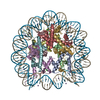
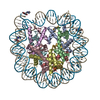
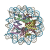
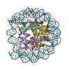
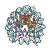
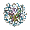
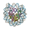
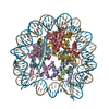
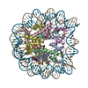
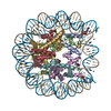
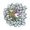
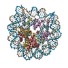
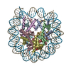
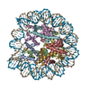
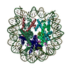
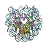
- PDB-1kx3: X-Ray Structure of the Nucleosome Core Particle, NCP146, at 2.0 A... -
+ Open data

- Basic information
| Entry | Database: PDB / ID: 1kx3 | ||||||
|---|---|---|---|---|---|---|---|
| Title | X-Ray Structure of the Nucleosome Core Particle, NCP146, at 2.0 A Resolution | ||||||
 Components Components |
| ||||||
 Keywords Keywords | STRUCTURAL PROTEIN/DNA / NUCLEOSOME / CHROMATIN / HISTONE / PROTEIN-DNA INTERACTION / NUCLEOPROTEIN / SUPERCOILED DNA / NUCLEOSOME CORE / PROTEIN-DNA COMPLEX / STRUCTURAL PROTEIN-DNA COMPLEX | ||||||
| Function / homology |  Function and homology information Function and homology informationstructural constituent of chromatin / nucleosome / nucleosome assembly / heterochromatin formation / protein heterodimerization activity / DNA binding / nucleoplasm / nucleus Similarity search - Function | ||||||
| Biological species |  Homo sapiens (human) Homo sapiens (human) | ||||||
| Method |  X-RAY DIFFRACTION / SYNCHROTRON / FOURIER SYNTHESIS / Resolution: 2 Å X-RAY DIFFRACTION / SYNCHROTRON / FOURIER SYNTHESIS / Resolution: 2 Å | ||||||
 Authors Authors | Davey, C.A. / Sargent, D.F. / Luger, K. / Maeder, A.W. / Richmond, T.J. | ||||||
 Citation Citation | Journal: J.Mol.Biol. / Year: 2002 Title: Solvent Mediated Interactions in the Structure of the Nucleosome Core Particle at 1.9 A Resolution Authors: Davey, C.A. / Sargent, D.F. / Luger, K. / Maeder, A.W. / Richmond, T.J. #1: Journal: Nature / Year: 1997Title: Crystal structure of the nucleosome core particle at 2.8 A resolution Authors: Luger, K. / Maeder, A.W. / Richmond, R.K. / Sargent, D.F. / Richmond, T.J. | ||||||
| History |
|
- Structure visualization
| Structure viewer | Molecule: MolmilJmol/JSmol |
|---|
- Downloads & links
-Download
| PDBx/mmCIF format | 1kx3.cif.gz | 346.8 KB | Display | PDBx/mmCIF format |
|---|---|---|---|---|
| PDB format | pdb1kx3.ent.gz | 265.1 KB | Display | PDB format |
| PDBx/mmJSON format | 1kx3.json.gz | Tree view | PDBx/mmJSON format | |
| Others |  Other downloads Other downloads |
-Validation report
| Arichive directory | https://data.pdbj.org/pub/pdb/validation_reports/kx/1kx3ftp://data.pdbj.org/pub/pdb/validation_reports/kx/1kx3 | HTTPS FTP |
|---|
-Related structure data
| Related structure data | 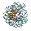 1kx4C  1kx5C  1aoiS S: Starting model for refinement C: citing same article ( |
|---|---|
| Similar structure data |


-Links
 PDBj
PDBj


- Assembly
| Deposited unit | 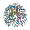
| ||||||||
|---|---|---|---|---|---|---|---|---|---|
| 1 |
| ||||||||
| Unit cell |
|
-Components
-DNA chain , 1 types, 2 molecules IJ
| #1: DNA chain | Mass: 45053.855 Da / Num. of mol.: 2 Source method: isolated from a genetically manipulated source Details: palindromic 146 base pair DNA duplex / Source: (gene. exp.) Homo sapiens (human)Description: DNA SEQUENCE SYNTHESIZED, CLONED, MULTIMERIZED, AND EXCISED FROM PLASMID Production host:  |
|---|
-Protein , 4 types, 8 molecules AEBFCGDH
| #2: Protein | Mass: 15303.930 Da / Num. of mol.: 2 Source method: isolated from a genetically manipulated source Source: (gene. exp.) #3: Protein | Mass: 11263.231 Da / Num. of mol.: 2 Source method: isolated from a genetically manipulated source Source: (gene. exp.) #4: Protein | Mass: 13907.163 Da / Num. of mol.: 2 Source method: isolated from a genetically manipulated source Source: (gene. exp.) #5: Protein | Mass: 13848.097 Da / Num. of mol.: 2 Source method: isolated from a genetically manipulated source Source: (gene. exp.) |
|---|
-Non-polymers , 2 types, 956 molecules 

| #6: Chemical | ChemComp-MN /  Mass: 54.938 Da / Num. of mol.: 13 / Source method: obtained synthetically / Formula: Mn Mass: 54.938 Da / Num. of mol.: 13 / Source method: obtained synthetically / Formula: Mn#7: Water | ChemComp-HOH / | Mass: 18.015 Da / Num. of mol.: 943 / Source method: isolated from a natural source / Formula: H2O |
|---|
-Experimental details
-Experiment
| Experiment | Method: X-RAY DIFFRACTION / Number of used crystals: 27 |
|---|
- Sample preparation
| Crystal | Density Matthews: 2.59 Å3/Da / Density % sol: 52.6 % | ||||||||||||||||||||||||||||||||||||||||||||||||||||||||
|---|---|---|---|---|---|---|---|---|---|---|---|---|---|---|---|---|---|---|---|---|---|---|---|---|---|---|---|---|---|---|---|---|---|---|---|---|---|---|---|---|---|---|---|---|---|---|---|---|---|---|---|---|---|---|---|---|---|
| Crystal grow | Temperature: 293 K / Method: vapor diffusion, sitting drop / pH: 6 Details: manganese chloride, potassium chloride, potassium cacodylate, pH 6.0, VAPOR DIFFUSION, SITTING DROP, temperature 293K | ||||||||||||||||||||||||||||||||||||||||||||||||||||||||
| Components of the solutions |
| ||||||||||||||||||||||||||||||||||||||||||||||||||||||||
| Crystal grow | *PLUS Method: vapor diffusion | ||||||||||||||||||||||||||||||||||||||||||||||||||||||||
| Components of the solutions | *PLUS
|
-Data collection
| Diffraction | Mean temperature: 103 K |
|---|---|
| Diffraction source | Source: SYNCHROTRON / Site: ESRF  / Beamline: ID09 / Wavelength: 0.85 Å / Beamline: ID09 / Wavelength: 0.85 Å |
| Detector | Type: MARRESEARCH / Detector: IMAGE PLATE / Date: Jun 2, 1996 |
| Radiation | Monochromator: Si 111 CHANNEL / Protocol: SINGLE WAVELENGTH / Monochromatic (M) / Laue (L): M / Scattering type: x-ray |
| Radiation wavelength | Wavelength: 0.85 Å / Relative weight: 1 |
| Reflection | Resolution: 1.97→99 Å / Num. all: 147830 / Num. obs: 145317 / % possible obs: 98.3 % / Observed criterion σ(F): 1 / Observed criterion σ(I): 1 / Redundancy: 7.4 % / Biso Wilson estimate: 35.7 Å2 / Rmerge(I) obs: 0.068 |
| Reflection shell | Resolution: 1.97→2.04 Å / Rmerge(I) obs: 0.495 / % possible all: 84.5 |
| Reflection | *PLUS Highest resolution: 2 Å / Lowest resolution: 99 Å |
| Reflection shell | *PLUS Highest resolution: 2 Å / % possible obs: 84.5 % |
- Processing
| Software |
| ||||||||||||||||||||||||||||||||||||
|---|---|---|---|---|---|---|---|---|---|---|---|---|---|---|---|---|---|---|---|---|---|---|---|---|---|---|---|---|---|---|---|---|---|---|---|---|---|
| Refinement | Method to determine structure: FOURIER SYNTHESIS Starting model: PDB entry 1AOI Resolution: 2→6 Å / Rfactor Rfree error: 0.005 / Data cutoff high absF: 952374.43 / Data cutoff low absF: 0 / Isotropic thermal model: RESTRAINED / Cross valid method: THROUGHOUT / σ(F): 0 / Stereochemistry target values: Engh & Huber
| ||||||||||||||||||||||||||||||||||||
| Displacement parameters | Biso mean: 58.2 Å2
| ||||||||||||||||||||||||||||||||||||
| Refine analyze |
| ||||||||||||||||||||||||||||||||||||
| Refinement step | Cycle: LAST / Resolution: 2→6 Å
| ||||||||||||||||||||||||||||||||||||
| Refine LS restraints |
| ||||||||||||||||||||||||||||||||||||
| LS refinement shell | Resolution: 2→2.12 Å / Rfactor Rfree error: 0.016 / Total num. of bins used: 6
| ||||||||||||||||||||||||||||||||||||
| Xplor file |
| ||||||||||||||||||||||||||||||||||||
| Refinement | *PLUS Highest resolution: 2 Å / Lowest resolution: 6 Å / Rfactor Rwork: 0.24 | ||||||||||||||||||||||||||||||||||||
| Solvent computation | *PLUS | ||||||||||||||||||||||||||||||||||||
| Displacement parameters | *PLUS | ||||||||||||||||||||||||||||||||||||
| Refine LS restraints | *PLUS
| ||||||||||||||||||||||||||||||||||||
| LS refinement shell | *PLUS Lowest resolution: 2.07 Å / Rfactor Rfree: 0.322 / Rfactor Rwork: 0.312 |
