 Movie
Movie Controller

[English] 日本語
 Yorodumi
- PDB-1zla: X-ray Structure of a Kaposi's sarcoma herpesvirus LANA peptide bo... -
+ Open data

- Basic information
| Entry | Database: PDB / ID: 1zla | ||||||
|---|---|---|---|---|---|---|---|
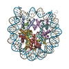
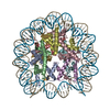
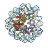
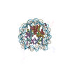
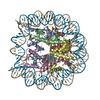
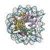
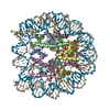
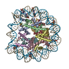
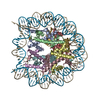
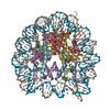
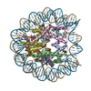
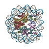
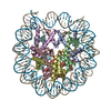
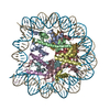
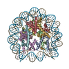
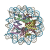
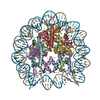
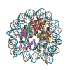
| Title | X-ray Structure of a Kaposi's sarcoma herpesvirus LANA peptide bound to the nucleosomal core | ||||||
 Components Components |
| ||||||
 Keywords Keywords | STRUCTURAL PROTEIN/DNA / Latency Associated Nuclear Antigen (LANA) / Kaposi's sarcoma Herpes Virus (KSHV) / Nucleosome core particle / chromatin / protein/protein interaction / STRUCTURAL PROTEIN-DNA complex | ||||||
| Function / homology |  Function and homology information Function and homology informationstructural constituent of chromatin / nucleosome / heterochromatin formation / nucleosome assembly / transcription coactivator activity / protein heterodimerization activity / host cell nucleus / positive regulation of transcription by RNA polymerase II / DNA binding / nucleoplasm / nucleus Similarity search - Function | ||||||
| Biological species |  Homo sapiens (human) Homo sapiens (human)Expression vector pET3-H2A (others) | ||||||
| Method |  X-RAY DIFFRACTION / MOLECULAR REPLACEMENT / Resolution: 2.9 Å X-RAY DIFFRACTION / MOLECULAR REPLACEMENT / Resolution: 2.9 Å | ||||||
 Authors Authors | Chodaparambil, J.V. / Barbera, A.J. / Kaye, K.M. / Luger, K. | ||||||
 Citation Citation | Journal: Science / Year: 2006 Title: The nucleosomal surface as a docking station for Kaposi's sarcoma herpesvirus LANA. Authors: Barbera, A.J. / Chodaparambil, J.V. / Kelley-Clarke, B. / Joukov, V. / Walter, J.C. / Luger, K. / Kaye, K.M. | ||||||
| History |
|
- Structure visualization
| Structure viewer | Molecule: MolmilJmol/JSmol |
|---|
- Downloads & links
-Download
| PDBx/mmCIF format | 1zla.cif.gz | 325.9 KB | Display | PDBx/mmCIF format |
|---|---|---|---|---|
| PDB format | pdb1zla.ent.gz | 246.3 KB | Display | PDB format |
| PDBx/mmJSON format | 1zla.json.gz | Tree view | PDBx/mmJSON format | |
| Others |  Other downloads Other downloads |
-Validation report
| Arichive directory | https://data.pdbj.org/pub/pdb/validation_reports/zl/1zlaftp://data.pdbj.org/pub/pdb/validation_reports/zl/1zla | HTTPS FTP |
|---|
-Related structure data
| Related structure data | 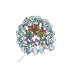 1aoiS S: Starting model for refinement |
|---|---|
| Similar structure data |


-Links
 PDBj
PDBj


- Assembly
| Deposited unit | 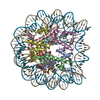
| ||||||||
|---|---|---|---|---|---|---|---|---|---|
| 1 |
| ||||||||
| Unit cell |
|
-Components
-Protein , 4 types, 8 molecules AEBFCGDH
| #2: Protein | Mass: 15340.973 Da / Num. of mol.: 2 Source method: isolated from a genetically manipulated source Source: (gene. exp.)  #3: Protein | Mass: 11263.231 Da / Num. of mol.: 2 Source method: isolated from a genetically manipulated source Source: (gene. exp.) #4: Protein | Mass: 14181.436 Da / Num. of mol.: 2 Source method: isolated from a genetically manipulated source Source: (gene. exp.) Expression vector pET3-H2A (others) / Gene: Histone H2A / Plasmid: pET / Production host: #5: Protein | Mass: 13794.008 Da / Num. of mol.: 2 Source method: isolated from a genetically manipulated source Source: (gene. exp.) |
|---|
-DNA chain / Protein/peptide / Non-polymers , 3 types, 69 molecules IJK
| #1: DNA chain | Mass: 45054.844 Da / Num. of mol.: 2 Source method: isolated from a genetically manipulated source Source: (gene. exp.) Homo sapiens (human) / Gene: Alpha-Satellite DNA / Plasmid: pUC / Production host: #6: Protein/peptide | | Mass: 2260.646 Da / Num. of mol.: 1 / Source method: obtained synthetically Details: N-terminal 1-23 amino acid region of Latency associated Nuclear Antigen (LANA)protein of Kaposi's Sarcoma Herpesvirus (KSHV) References: GenBank: 5669894, UniProt: Q9DUM3*PLUS #7: Water | ChemComp-HOH / | Mass: 18.015 Da / Num. of mol.: 66 / Source method: isolated from a natural source / Formula: H2O |
|---|
-Experimental details
-Experiment
| Experiment | Method: X-RAY DIFFRACTION / Number of used crystals: 1 |
|---|
- Sample preparation
| Crystal | Density Matthews: 2.59 Å3/Da / Density % sol: 52.46 % | ||||||||||||||||||||||||
|---|---|---|---|---|---|---|---|---|---|---|---|---|---|---|---|---|---|---|---|---|---|---|---|---|---|
| Crystal grow | Temperature: 292 K / Method: vapor diffusion, sitting drop / pH: 6 Details: Manganese chloride, Potassium chloride, Potassium cacodylate, pH 6.0, VAPOR DIFFUSION, SITTING DROP, temperature 292K | ||||||||||||||||||||||||
| Components of the solutions |
|
-Data collection
| Diffraction | Mean temperature: 100 K |
|---|---|
| Diffraction source | Source: ROTATING ANODE / Type: RIGAKU / Wavelength: 1.5418 Å |
| Detector | Type: RIGAKU RAXIS IV / Detector: IMAGE PLATE / Date: Jan 15, 2005 / Details: mirrors |
| Radiation | Monochromator: Cu / Protocol: SINGLE WAVELENGTH / Monochromatic (M) / Laue (L): M / Scattering type: x-ray |
| Radiation wavelength | Wavelength: 1.5418 Å / Relative weight: 1 |
| Reflection | Resolution: 2.9→50 Å / Num. all: 48638 / Num. obs: 47855 / % possible obs: 90.8 % / Observed criterion σ(F): 0 / Observed criterion σ(I): 15273.7 / Redundancy: 2.66 % / Rmerge(I) obs: 0.045 / Net I/σ(I): 14.99 |
| Reflection shell | Resolution: 2.9→2.97 Å / Rmerge(I) obs: 0.239 / Mean I/σ(I) obs: 3.64 / % possible all: 82.8 |
- Processing
| Software |
| |||||||||||||||||||||||||
|---|---|---|---|---|---|---|---|---|---|---|---|---|---|---|---|---|---|---|---|---|---|---|---|---|---|---|
| Refinement | Method to determine structure: MOLECULAR REPLACEMENT Starting model: PDB entry 1aoi Resolution: 2.9→50 Å / Cross valid method: Thorughout / σ(F): 0 / σ(I): 0 / Stereochemistry target values: Engh & Huber
| |||||||||||||||||||||||||
| Displacement parameters |
| |||||||||||||||||||||||||
| Refinement step | Cycle: LAST / Resolution: 2.9→50 Å
| |||||||||||||||||||||||||
| Refine LS restraints |
| |||||||||||||||||||||||||
| Xplor file |
|
