 Movie
Movie Controller


+ Open data

- Basic information
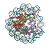
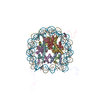
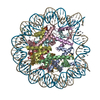
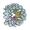
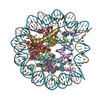
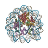
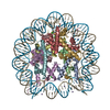
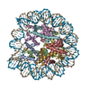
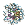
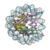
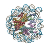
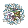
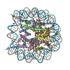
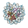
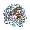
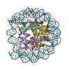
| Entry | Database: PDB / ID: 2nzd | ||||||
|---|---|---|---|---|---|---|---|
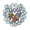
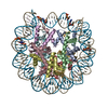
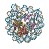
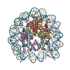
| Title | Nucleosome core particle containing 145 bp of DNA | ||||||
 Components Components |
| ||||||
 Keywords Keywords | STRUCTURAL PROTEIN/DNA / nucleosome / chromatin / histone / DNA stretching / DNA kinking / double-helix / STRUCTURAL PROTEIN-DNA COMPLEX | ||||||
| Function / homology |  Function and homology information Function and homology informationstructural constituent of chromatin / nucleosome / nucleosome assembly / heterochromatin formation / protein heterodimerization activity / DNA binding / nucleoplasm / nucleus Similarity search - Function | ||||||
| Biological species | |||||||
| Method |  X-RAY DIFFRACTION / FOURIER SYNTHESIS / Resolution: 2.65 Å X-RAY DIFFRACTION / FOURIER SYNTHESIS / Resolution: 2.65 Å | ||||||
 Authors Authors | Ong, M.S. / Richmond, T.J. / Davey, C.A. | ||||||
 Citation Citation | Journal: J.Mol.Biol. / Year: 2007 Title: DNA stretching and extreme kinking in the nucleosome core Authors: Ong, M.S. / Richmond, T.J. / Davey, C.A. | ||||||
| History |
|
- Structure visualization
| Structure viewer | Molecule: MolmilJmol/JSmol |
|---|
- Downloads & links
-Download
| PDBx/mmCIF format | 2nzd.cif.gz | 325.4 KB | Display | PDBx/mmCIF format |
|---|---|---|---|---|
| PDB format | pdb2nzd.ent.gz | 245.5 KB | Display | PDB format |
| PDBx/mmJSON format | 2nzd.json.gz | Tree view | PDBx/mmJSON format | |
| Others |  Other downloads Other downloads |
-Validation report
| Arichive directory | https://data.pdbj.org/pub/pdb/validation_reports/nz/2nzdftp://data.pdbj.org/pub/pdb/validation_reports/nz/2nzd | HTTPS FTP |
|---|
-Related structure data
| Related structure data | 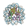 1kx3S S: Starting model for refinement |
|---|---|
| Similar structure data |


-Links
 PDBj
PDBj


- Assembly
| Deposited unit | 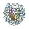
| ||||||||
|---|---|---|---|---|---|---|---|---|---|
| 1 |
| ||||||||
| Unit cell |
|
-Components
-DNA chain , 2 types, 2 molecules IJ
| #1: DNA chain | Mass: 44749.664 Da / Num. of mol.: 1 / Source method: obtained synthetically |
|---|---|
| #2: DNA chain | Mass: 44740.648 Da / Num. of mol.: 1 / Source method: obtained synthetically |
-Protein , 4 types, 8 molecules AEBFCGDH
| #3: Protein | Mass: 15303.930 Da / Num. of mol.: 2 Source method: isolated from a genetically manipulated source Source: (gene. exp.)  #4: Protein | Mass: 11263.231 Da / Num. of mol.: 2 Source method: isolated from a genetically manipulated source Source: (gene. exp.) #5: Protein | Mass: 12941.095 Da / Num. of mol.: 2 Source method: isolated from a genetically manipulated source Source: (gene. exp.) #6: Protein | Mass: 13848.097 Da / Num. of mol.: 2 Source method: isolated from a genetically manipulated source Source: (gene. exp.) |
|---|
-Non-polymers , 2 types, 133 molecules 

| #7: Chemical | ChemComp-MN /  Mass: 54.938 Da / Num. of mol.: 11 / Source method: obtained synthetically / Formula: Mn Mass: 54.938 Da / Num. of mol.: 11 / Source method: obtained synthetically / Formula: Mn#8: Water | ChemComp-HOH / | Mass: 18.015 Da / Num. of mol.: 122 / Source method: isolated from a natural source / Formula: H2O |
|---|
-Experimental details
-Experiment
| Experiment | Method: X-RAY DIFFRACTION / Number of used crystals: 1 |
|---|
- Sample preparation
| Crystal | Density Matthews: 2.92 Å3/Da / Density % sol: 57.86 % | ||||||||||||||||||||||||||||||||||||
|---|---|---|---|---|---|---|---|---|---|---|---|---|---|---|---|---|---|---|---|---|---|---|---|---|---|---|---|---|---|---|---|---|---|---|---|---|---|
| Crystal grow | Temperature: 291 K / Method: vapor diffusion, hanging drop / pH: 6 Details: 85 mM MnCl2, 60 mM KCl, 20 mM K-Cacodylate, 4 mg/ml NCP over well with 1/2 conc., pH 6.0, VAPOR DIFFUSION, HANGING DROP, temperature 291K | ||||||||||||||||||||||||||||||||||||
| Components of the solutions |
|
-Data collection
| Diffraction | Mean temperature: 98 K |
|---|---|
| Diffraction source | Source: ROTATING ANODE / Type: RIGAKU / Wavelength: 1.542 Å |
| Detector | Type: RIGAKU RAXIS IV / Detector: IMAGE PLATE / Date: Sep 5, 2006 / Details: osmic mirrors |
| Radiation | Monochromator: graphite / Protocol: SINGLE WAVELENGTH / Monochromatic (M) / Laue (L): M / Scattering type: x-ray |
| Radiation wavelength | Wavelength: 1.542 Å / Relative weight: 1 |
| Reflection | Resolution: 2.65→50.7 Å / Num. all: 61751 / Num. obs: 56193 / % possible obs: 91 % / Observed criterion σ(F): 0 / Observed criterion σ(I): 0 / Redundancy: 6.3 % / Rmerge(I) obs: 0.065 / Rsym value: 0.065 / Net I/σ(I): 20.9 |
| Reflection shell | Resolution: 2.65→2.79 Å / Redundancy: 4.9 % / Rmerge(I) obs: 0.48 / Mean I/σ(I) obs: 2.7 / Num. measured all: 27467 / Num. unique all: 5579 / Rsym value: 0.48 / % possible all: 63.9 |
- Processing
| Software |
| ||||||||||||||||||||||||||||||||||||||||||||||||||||||||||||||||||||||||||||||||||||||||||
|---|---|---|---|---|---|---|---|---|---|---|---|---|---|---|---|---|---|---|---|---|---|---|---|---|---|---|---|---|---|---|---|---|---|---|---|---|---|---|---|---|---|---|---|---|---|---|---|---|---|---|---|---|---|---|---|---|---|---|---|---|---|---|---|---|---|---|---|---|---|---|---|---|---|---|---|---|---|---|---|---|---|---|---|---|---|---|---|---|---|---|---|
| Refinement | Method to determine structure: FOURIER SYNTHESIS Starting model: PDB ENTRY 1KX3 Resolution: 2.65→40 Å / Cor.coef. Fo:Fc: 0.921 / Cor.coef. Fo:Fc free: 0.892 / SU B: 11.836 / SU ML: 0.259 / Cross valid method: THROUGHOUT / σ(F): 0 / ESU R: 0.887 / ESU R Free: 0.367 / Stereochemistry target values: MAXIMUM LIKELIHOOD / Details: HYDROGENS HAVE BEEN ADDED IN THE RIDING POSITIONS
| ||||||||||||||||||||||||||||||||||||||||||||||||||||||||||||||||||||||||||||||||||||||||||
| Solvent computation | Ion probe radii: 0.8 Å / Shrinkage radii: 0.8 Å / VDW probe radii: 1.4 Å / Solvent model: MASK | ||||||||||||||||||||||||||||||||||||||||||||||||||||||||||||||||||||||||||||||||||||||||||
| Displacement parameters | Biso mean: 64.218 Å2
| ||||||||||||||||||||||||||||||||||||||||||||||||||||||||||||||||||||||||||||||||||||||||||
| Refine analyze | Luzzati coordinate error free: 0.37 Å | ||||||||||||||||||||||||||||||||||||||||||||||||||||||||||||||||||||||||||||||||||||||||||
| Refinement step | Cycle: LAST / Resolution: 2.65→40 Å
| ||||||||||||||||||||||||||||||||||||||||||||||||||||||||||||||||||||||||||||||||||||||||||
| Refine LS restraints |
| ||||||||||||||||||||||||||||||||||||||||||||||||||||||||||||||||||||||||||||||||||||||||||
| LS refinement shell | Resolution: 2.65→2.719 Å / Total num. of bins used: 20
|
