 Movie
Movie Controller


+ Open data

- Basic information
| Entry | Database: PDB / ID: 5f99 | ||||||
|---|---|---|---|---|---|---|---|
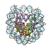
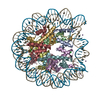
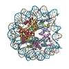
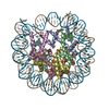
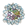
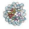
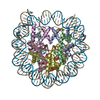
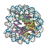
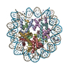
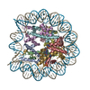
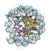
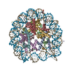
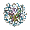
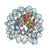
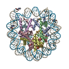
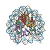
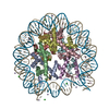
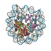
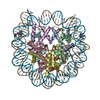
| Title | X-ray Structure of the MMTV-A Nucleosome Core Particle | ||||||
 Components Components |
| ||||||
 Keywords Keywords | DNA BINDING PROTEIN / nucleosome core particle histone DNA | ||||||
| Function / homology |  Function and homology information Function and homology informationstructural constituent of chromatin / nucleosome / nucleosome assembly / heterochromatin formation / protein heterodimerization activity / DNA binding / nucleoplasm / nucleus Similarity search - Function | ||||||
| Biological species |  Mouse mammary tumor virus Mouse mammary tumor virus | ||||||
| Method |  X-RAY DIFFRACTION / SYNCHROTRON / MOLECULAR REPLACEMENT / Resolution: 2.63 Å X-RAY DIFFRACTION / SYNCHROTRON / MOLECULAR REPLACEMENT / Resolution: 2.63 Å | ||||||
 Authors Authors | Frouws, T.D. / Richmond, T.J. | ||||||
 Citation Citation | Journal: Proc.Natl.Acad.Sci.USA / Year: 2016 Title: X-ray structure of the MMTV-A nucleosome core. Authors: Frouws, T.D. / Duda, S.C. / Richmond, T.J. | ||||||
| History |
|
- Structure visualization
| Structure viewer | Molecule: MolmilJmol/JSmol |
|---|
- Downloads & links
-Download
| PDBx/mmCIF format | 5f99.cif.gz | 361 KB | Display | PDBx/mmCIF format |
|---|---|---|---|---|
| PDB format | pdb5f99.ent.gz | 274 KB | Display | PDB format |
| PDBx/mmJSON format | 5f99.json.gz | Tree view | PDBx/mmJSON format | |
| Others |  Other downloads Other downloads |
-Validation report
| Arichive directory | https://data.pdbj.org/pub/pdb/validation_reports/f9/5f99ftp://data.pdbj.org/pub/pdb/validation_reports/f9/5f99 | HTTPS FTP |
|---|
-Related structure data
| Related structure data | 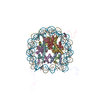 1kx5S S: Starting model for refinement |
|---|---|
| Similar structure data |

-Links
 PDBj
PDBj


- Assembly
| Deposited unit | 
| ||||||||
|---|---|---|---|---|---|---|---|---|---|
| 1 |
| ||||||||
| Unit cell |
|
-Components
-Protein , 4 types, 8 molecules AEBFCGDH
| #1: Protein | Mass: 15271.863 Da / Num. of mol.: 2 / Mutation: C110A Source method: isolated from a genetically manipulated source Source: (gene. exp.)  #2: Protein | Mass: 11413.473 Da / Num. of mol.: 2 / Mutation: H18R Source method: isolated from a genetically manipulated source Source: (gene. exp.) #3: Protein | Mass: 13978.241 Da / Num. of mol.: 2 Source method: isolated from a genetically manipulated source Source: (gene. exp.) #4: Protein | Mass: 13524.752 Da / Num. of mol.: 2 Source method: isolated from a genetically manipulated source Source: (gene. exp.) |
|---|
-DNA chain , 2 types, 2 molecules IJ
| #5: DNA chain | Mass: 44849.496 Da / Num. of mol.: 1 Source method: isolated from a genetically manipulated source Source: (gene. exp.) Mouse mammary tumor virus / Plasmid: pUC57 / Production host: |
|---|---|
| #6: DNA chain | Mass: 45901.312 Da / Num. of mol.: 1 Source method: isolated from a genetically manipulated source Source: (gene. exp.) Mouse mammary tumor virus / Plasmid: pUC57 / Production host: |
-Non-polymers , 3 types, 941 molecules 


| #7: Chemical | ChemComp-CL /  Mass: 35.453 Da / Num. of mol.: 4 / Source method: obtained synthetically / Formula: Cl Mass: 35.453 Da / Num. of mol.: 4 / Source method: obtained synthetically / Formula: Cl#8: Chemical | ChemComp-MG / |  Mass: 24.305 Da / Num. of mol.: 1 / Source method: obtained synthetically / Formula: Mg Mass: 24.305 Da / Num. of mol.: 1 / Source method: obtained synthetically / Formula: Mg#9: Water | ChemComp-HOH / | Mass: 18.015 Da / Num. of mol.: 936 / Source method: isolated from a natural source / Formula: H2O |
|---|
-Experimental details
-Experiment
| Experiment | Method: X-RAY DIFFRACTION |
|---|
- Sample preparation
| Crystal | Density Matthews: 2.64 Å3/Da / Density % sol: 53.46 % / Description: Hollow hexagonal rods |
|---|---|
| Crystal grow | Temperature: 295 K / Method: vapor diffusion, sitting drop / pH: 6 Details: sample was mixed 1:1 with 10 mM K-cacodylate, pH 6.0, 180 mM MgCl2, 50 mM KCl and equilibrated against a 1:4 dilution of the same solution Temp details: Rumed incubator |
-Data collection
| Diffraction | Mean temperature: 90 K |
|---|---|
| Diffraction source | Source: SYNCHROTRON / Site: SLS  / Beamline: X06SA / Wavelength: 1 Å / Beamline: X06SA / Wavelength: 1 Å |
| Detector | Type: DECTRIS PILATUS 6M / Detector: PIXEL / Date: Oct 28, 2012 |
| Radiation | Protocol: SINGLE WAVELENGTH / Monochromatic (M) / Laue (L): M / Scattering type: x-ray |
| Radiation wavelength | Wavelength: 1 Å / Relative weight: 1 |
| Reflection | Resolution: 2.63→29.68 Å / Num. obs: 59659 / % possible obs: 94.2 % / Redundancy: 6.8 % / Rmerge(I) obs: 0.083 / Net I/σ(I): 14.2 |
| Reflection shell | Resolution: 2.63→2.78 Å / Redundancy: 2.5 % / Rmerge(I) obs: 0.333 / Mean I/σ(I) obs: 1.8 / % possible all: 73.8 |
- Processing
| Software |
| ||||||||||||||||||||
|---|---|---|---|---|---|---|---|---|---|---|---|---|---|---|---|---|---|---|---|---|---|
| Refinement | Method to determine structure: MOLECULAR REPLACEMENT Starting model: 1KX5 Resolution: 2.63→29.68 Å / Cross valid method: FREE R-VALUE
| ||||||||||||||||||||
| Refinement step | Cycle: LAST / Resolution: 2.63→29.68 Å
|
