- PDB-1yd9: 1.6A Crystal Structure of the Non-Histone Domain of the Histone V... -
+
Open data
ID or keywords:
Loading...
-
Basic information
Entry
Database: PDB / ID: 1yd9
Title
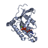
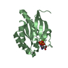
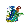
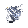
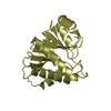
1.6A Crystal Structure of the Non-Histone Domain of the Histone Variant MacroH2A1.1.
Components
Core histone macro-H2A.1
Keywords
STRUCTURAL PROTEIN / Alpha-Beta Structure / A1pp Domain / Macro-Domain
Function / homology
Function and homology information
negative regulation of cell cycle G2/M phase transition / positive regulation of endodermal cell differentiation / negative regulation of protein localization to chromosome, telomeric region / regulation of NAD metabolic process / positive regulation of response to oxidative stress / Barr body / positive regulation of maintenance of mitotic sister chromatid cohesion / regulation of response to oxidative stress / ADP-D-ribose binding / ADP-D-ribose modification-dependent protein binding ...negative regulation of cell cycle G2/M phase transition / positive regulation of endodermal cell differentiation / negative regulation of protein localization to chromosome, telomeric region / regulation of NAD metabolic process / positive regulation of response to oxidative stress / Barr body / positive regulation of maintenance of mitotic sister chromatid cohesion / regulation of response to oxidative stress / ADP-D-ribose binding / ADP-D-ribose modification-dependent protein binding / negative regulation of transcription of nucleolar large rRNA by RNA polymerase I / double-stranded methylated DNA binding / regulation of oxidative phosphorylation / rDNA binding / positive regulation of keratinocyte differentiation / establishment of protein localization to chromatin / dosage compensation by inactivation of X chromosome / negative regulation of response to oxidative stress / nucleosomal DNA binding / nuclear chromosome / negative regulation of gene expression, epigenetic / regulation of lipid metabolic process / protein serine/threonine kinase inhibitor activity / pericentric heterochromatin / site of DNA damage / condensed chromosome / epigenetic regulation of gene expression / transcription initiation-coupled chromatin remodeling / RNA polymerase II transcription regulatory region sequence-specific DNA binding / promoter-specific chromatin binding / chromatin DNA binding / structural constituent of chromatin / nucleosome / nucleosome assembly / heterochromatin formation / transcription cis-regulatory region binding / protein heterodimerization activity / DNA repair / chromatin binding / protein kinase binding / nucleolus / chromatin / enzyme binding / negative regulation of transcription by RNA polymerase II / nucleoplasm / nucleus Similarity search - Function
SEQUENCE The first five residues GPLGS are cloning artifacts. The sequence 6-51 is variance ...SEQUENCE The first five residues GPLGS are cloning artifacts. The sequence 6-51 is variance splicing in isoform 1.
In the structure databanks used in Yorodumi, some data are registered as the other names, "COVID-19 virus" and "2019-nCoV". Here are the details of the virus and the list of structure data.
Jan 31, 2019. EMDB accession codes are about to change! (news from PDBe EMDB page)
EMDB accession codes are about to change! (news from PDBe EMDB page)
The allocation of 4 digits for EMDB accession codes will soon come to an end. Whilst these codes will remain in use, new EMDB accession codes will include an additional digit and will expand incrementally as the available range of codes is exhausted. The current 4-digit format prefixed with “EMD-” (i.e. EMD-XXXX) will advance to a 5-digit format (i.e. EMD-XXXXX), and so on. It is currently estimated that the 4-digit codes will be depleted around Spring 2019, at which point the 5-digit format will come into force.
The EM Navigator/Yorodumi systems omit the EMD- prefix.
Related info.:Q: What is EMD? / ID/Accession-code notation in Yorodumi/EM Navigator
Yorodumi is a browser for structure data from EMDB, PDB, SASBDB, etc.
This page is also the successor to EM Navigator detail page, and also detail information page/front-end page for Omokage search.
The word "yorodu" (or yorozu) is an old Japanese word meaning "ten thousand". "mi" (miru) is to see.
Related info.:EMDB / PDB / SASBDB / Comparison of 3 databanks / Yorodumi Search / Aug 31, 2016. New EM Navigator & Yorodumi / Yorodumi Papers / Jmol/JSmol / Function and homology information / Changes in new EM Navigator and Yorodumi
 Movie
Movie Controller

 Yorodumi
 Open data
 Basic information
 Components
 Keywords
 Function and homology information
 X-RAY DIFFRACTION /
  Authors
 Citation
 Structure visualization
 Downloads & links
 Other downloads
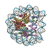


 PDBj
PDBj


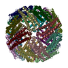
 Assembly
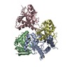


 Mass: 196.967 Da / Num. of mol.: 4 / Source method: obtained synthetically / Formula: Au
 Mass: 18.015 Da / Num. of mol.: 540 / Source method: isolated from a natural source / Formula: H2O
 Sample preparation
 Processing