 Movie
Movie Controller


+ Open data

- Basic information
| Entry | Database: PDB / ID: 2nqb | ||||||
|---|---|---|---|---|---|---|---|
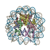
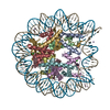
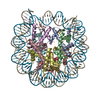
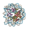
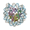
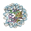
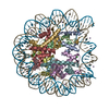
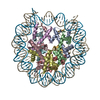
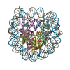
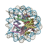
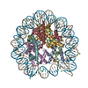
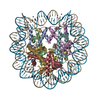
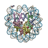
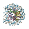
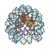
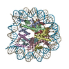
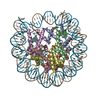
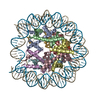
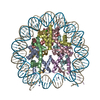
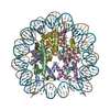
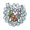
| Title | Drosophila Nucleosome Structure | ||||||
 Components Components |
| ||||||
 Keywords Keywords | STRUCTURAL PROTEIN/DNA / Nucleosome / NCP / chromatin / histone / STRUCTURAL PROTEIN-DNA COMPLEX | ||||||
| Function / homology |  Function and homology information Function and homology informationHDMs demethylate histones / PKMTs methylate histone lysines / Interleukin-7 signaling / Chromatin modifying enzymes / SUMOylation of chromatin organization proteins / Recruitment and ATM-mediated phosphorylation of repair and signaling proteins at DNA double strand breaks / RMTs methylate histone arginines / E3 ubiquitin ligases ubiquitinate target proteins / RCAF complex / Factors involved in megakaryocyte development and platelet production ...HDMs demethylate histones / PKMTs methylate histone lysines / Interleukin-7 signaling / Chromatin modifying enzymes / SUMOylation of chromatin organization proteins / Recruitment and ATM-mediated phosphorylation of repair and signaling proteins at DNA double strand breaks / RMTs methylate histone arginines / E3 ubiquitin ligases ubiquitinate target proteins / RCAF complex / Factors involved in megakaryocyte development and platelet production / SIRT1 negatively regulates rRNA expression / NoRC negatively regulates rRNA expression / Activated PKN1 stimulates transcription of AR (androgen receptor) regulated genes KLK2 and KLK3 / polytene chromosome band / PRC2 methylates histones and DNA / Ub-specific processing proteases / Negative Regulation of CDH1 Gene Transcription / MLL4 and MLL3 complexes regulate expression of PPARG target genes in adipogenesis and hepatic steatosis / Regulation of PD-L1(CD274) transcription / larval somatic muscle development / RNA Polymerase I Promoter Escape / Regulation of endogenous retroelements by KRAB-ZFP proteins / RUNX1 regulates genes involved in megakaryocyte differentiation and platelet function / Senescence-Associated Secretory Phenotype (SASP) / Transcriptional regulation by small RNAs / Estrogen-dependent gene expression / HATs acetylate histones / Assembly of the ORC complex at the origin of replication / Oxidative Stress Induced Senescence / polytene chromosome / nuclear chromosome / nucleosomal DNA binding / structural constituent of chromatin / nucleosome / nucleosome assembly / chromosome / chromatin organization / heterochromatin formation / protein heterodimerization activity / chromatin / protein-containing complex binding / DNA binding / nucleus Similarity search - Function | ||||||
| Biological species |  Homo sapiens (human) Homo sapiens (human) | ||||||
| Method |  X-RAY DIFFRACTION / SYNCHROTRON / MOLECULAR REPLACEMENT / Resolution: 2.3 Å X-RAY DIFFRACTION / SYNCHROTRON / MOLECULAR REPLACEMENT / Resolution: 2.3 Å | ||||||
 Authors Authors | Luger, K. / Chakravarthy, S. | ||||||
 Citation Citation | Journal: To be Published Title: Comparative analysis of nucleosome structures from different species. Authors: Chakravarthy, S. / Luger, K. | ||||||
| History |
|
- Structure visualization
| Structure viewer | Molecule: MolmilJmol/JSmol |
|---|
- Downloads & links
-Download
| PDBx/mmCIF format | 2nqb.cif.gz | 327.2 KB | Display | PDBx/mmCIF format |
|---|---|---|---|---|
| PDB format | pdb2nqb.ent.gz | 249.4 KB | Display | PDB format |
| PDBx/mmJSON format | 2nqb.json.gz | Tree view | PDBx/mmJSON format | |
| Others |  Other downloads Other downloads |
-Validation report
| Arichive directory | https://data.pdbj.org/pub/pdb/validation_reports/nq/2nqbftp://data.pdbj.org/pub/pdb/validation_reports/nq/2nqb | HTTPS FTP |
|---|
-Related structure data
| Related structure data | 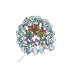 1aoiS S: Starting model for refinement |
|---|---|
| Similar structure data |

-Links
 PDBj
PDBj


- Assembly
| Deposited unit | 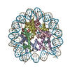
| ||||||||
|---|---|---|---|---|---|---|---|---|---|
| 1 |
| ||||||||
| Unit cell |
| ||||||||
| Details | Each nucleosome has two copies each of histones H2A, H2B, H3 and H4, and a 146 base pairs long palindromic strand of DNA derived from human alpha-satellite sequence. |
-Components
-Protein , 4 types, 8 molecules AEBFCGDH
| #2: Protein | Mass: 15289.904 Da / Num. of mol.: 2 Source method: isolated from a genetically manipulated source Source: (gene. exp.)  #3: Protein | Mass: 11390.414 Da / Num. of mol.: 2 Source method: isolated from a genetically manipulated source Source: (gene. exp.) #4: Protein | Mass: 13257.529 Da / Num. of mol.: 2 Source method: isolated from a genetically manipulated source Source: (gene. exp.) #5: Protein | Mass: 13680.951 Da / Num. of mol.: 2 Source method: isolated from a genetically manipulated source Source: (gene. exp.) |
|---|
-DNA chain / Non-polymers , 2 types, 266 molecules IJ
| #1: DNA chain | Mass: 45054.844 Da / Num. of mol.: 2 Source method: isolated from a genetically manipulated source Source: (gene. exp.) Homo sapiens (human) / Gene: His3 / Production host: #6: Water | ChemComp-HOH / | Mass: 18.015 Da / Num. of mol.: 264 / Source method: isolated from a natural source / Formula: H2O |
|---|
-Experimental details
-Experiment
| Experiment | Method: X-RAY DIFFRACTION / Number of used crystals: 1 |
|---|
- Sample preparation
| Crystal | Density Matthews: 2.64 Å3/Da / Density % sol: 53.37 % | ||||||||||||||||||||||||||||||||||||
|---|---|---|---|---|---|---|---|---|---|---|---|---|---|---|---|---|---|---|---|---|---|---|---|---|---|---|---|---|---|---|---|---|---|---|---|---|---|
| Crystal grow | Temperature: 292 K / Method: vapor diffusion, sitting drop / pH: 6 Details: Constituents of the crystallization buffer: Potassium Chloride, Manganese Chloride, and Potassium Cacodylate., pH 6.0, VAPOR DIFFUSION, SITTING DROP, temperature 292K | ||||||||||||||||||||||||||||||||||||
| Components of the solutions |
|
-Data collection
| Diffraction | Mean temperature: 93 K |
|---|---|
| Diffraction source | Source: SYNCHROTRON / Site: ALS  / Beamline: 5.0.2 / Wavelength: 1.1 Å / Beamline: 5.0.2 / Wavelength: 1.1 Å |
| Radiation | Protocol: SINGLE WAVELENGTH / Monochromatic (M) / Laue (L): M / Scattering type: x-ray |
| Radiation wavelength | Wavelength: 1.1 Å / Relative weight: 1 |
| Reflection | Resolution: 2.3→99 Å / Num. all: 94877 / Num. obs: 93949 / % possible obs: 98.2 % / Rmerge(I) obs: 0.045 / Net I/σ(I): 22 |
| Reflection shell | Resolution: 2.3→2.35 Å / Rmerge(I) obs: 0.302 / Mean I/σ(I) obs: 3 / Num. unique all: 5358 / % possible all: 85 |
- Processing
| Software |
| ||||||||||||||||||||
|---|---|---|---|---|---|---|---|---|---|---|---|---|---|---|---|---|---|---|---|---|---|
| Refinement | Method to determine structure: MOLECULAR REPLACEMENT Starting model: PDB entry 1AOI Resolution: 2.3→99 Å
| ||||||||||||||||||||
| Displacement parameters |
| ||||||||||||||||||||
| Refinement step | Cycle: LAST / Resolution: 2.3→99 Å
| ||||||||||||||||||||
| Refine LS restraints |
| ||||||||||||||||||||
| Xplor file |
|
