 Movie
Movie Controller

[English] 日本語
 Yorodumi
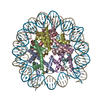
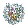
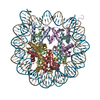
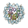
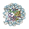
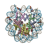
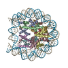
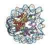
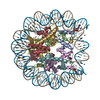
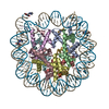
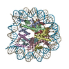
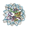
- PDB-4xzq: Nucleosome disassembly by RSC and SWI/SNF is enhanced by H3 acety... -
+ Open data

- Basic information
| Entry | Database: PDB / ID: 4xzq | ||||||||||||
|---|---|---|---|---|---|---|---|---|---|---|---|---|---|
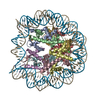
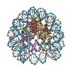
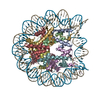
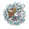
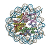
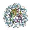
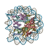
| Title | Nucleosome disassembly by RSC and SWI/SNF is enhanced by H3 acetylation near the nucleosome dyad axis | ||||||||||||
 Components Components |
| ||||||||||||
 Keywords Keywords | STRUCTURAL PROTEIN/DNA / dyad axis / STRUCTURAL PROTEIN-DNA complex | ||||||||||||
| Function / homology |  Function and homology information Function and homology informationstructural constituent of chromatin / nucleosome / nucleosome assembly / heterochromatin formation / protein heterodimerization activity / DNA binding / nucleoplasm / nucleus Similarity search - Function | ||||||||||||
| Biological species |  Homo sapiens (human) Homo sapiens (human) | ||||||||||||
| Method |  X-RAY DIFFRACTION / SYNCHROTRON / MOLECULAR REPLACEMENT / Resolution: 2.4 Å X-RAY DIFFRACTION / SYNCHROTRON / MOLECULAR REPLACEMENT / Resolution: 2.4 Å | ||||||||||||
 Authors Authors | Dechassa, M.L. / Luger, K. / Chatterjee, N. / North, J.A. / Manohar, M. / Prasad, R. / Ottessen, J.J. / Poirier, M.G. / Bartholomew, B. | ||||||||||||
| Funding support |  United States, 3items United States, 3items
| ||||||||||||
 Citation Citation | Journal: Mol.Cell.Biol. / Year: 2015 Title: Histone Acetylation near the Nucleosome Dyad Axis Enhances Nucleosome Disassembly by RSC and SWI/SNF. Authors: Chatterjee, N. / North, J.A. / Dechassa, M.L. / Manohar, M. / Prasad, R. / Luger, K. / Ottesen, J.J. / Poirier, M.G. / Bartholomew, B. | ||||||||||||
| History |
|
- Structure visualization
| Structure viewer | Molecule: MolmilJmol/JSmol |
|---|
- Downloads & links
-Download
| PDBx/mmCIF format | 4xzq.cif.gz | 320.4 KB | Display | PDBx/mmCIF format |
|---|---|---|---|---|
| PDB format | pdb4xzq.ent.gz | 244.1 KB | Display | PDB format |
| PDBx/mmJSON format | 4xzq.json.gz | Tree view | PDBx/mmJSON format | |
| Others |  Other downloads Other downloads |
-Validation report
| Arichive directory | https://data.pdbj.org/pub/pdb/validation_reports/xz/4xzqftp://data.pdbj.org/pub/pdb/validation_reports/xz/4xzq | HTTPS FTP |
|---|
-Related structure data
| Related structure data | 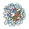 4ys3C 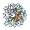 4z66C  1p3lS S: Starting model for refinement C: citing same article ( |
|---|---|
| Similar structure data |

-Links
 PDBj
PDBj


- Assembly
| Deposited unit | 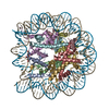
| ||||||||
|---|---|---|---|---|---|---|---|---|---|
| 1 |
| ||||||||
| Unit cell |
|
-Components
-Protein , 4 types, 8 molecules AEBFCGDH
| #1: Protein | Mass: 11543.464 Da / Num. of mol.: 2 / Fragment: residues 39-136 Source method: isolated from a genetically manipulated source Details: H3 acetylated at lysine 115 / Source: (gene. exp.)  #2: Protein | Mass: 8910.394 Da / Num. of mol.: 2 / Fragment: residues 25-103 Source method: isolated from a genetically manipulated source Source: (gene. exp.) #3: Protein | Mass: 11724.677 Da / Num. of mol.: 2 / Fragment: residues 15-121 Source method: isolated from a genetically manipulated source Source: (gene. exp.) #4: Protein | Mass: 10376.928 Da / Num. of mol.: 2 / Fragment: residues 34-126 Source method: isolated from a genetically manipulated source Source: (gene. exp.) |
|---|
-DNA chain , 2 types, 2 molecules IJ
| #5: DNA chain | Mass: 45368.051 Da / Num. of mol.: 1 / Source method: obtained synthetically / Source: (synth.) Homo sapiens (human) |
|---|---|
| #6: DNA chain | Mass: 45359.035 Da / Num. of mol.: 1 / Source method: obtained synthetically / Source: (synth.) Homo sapiens (human) |
-Non-polymers , 1 types, 99 molecules 
| #7: Water | ChemComp-HOH / Mass: 18.015 Da / Num. of mol.: 99 / Source method: isolated from a natural source / Formula: H2O |
|---|
-Details
| Has protein modification | Y |
|---|
-Experimental details
-Experiment
| Experiment | Method: X-RAY DIFFRACTION / Number of used crystals: 1 |
|---|
- Sample preparation
| Crystal | Density Matthews: 2.97 Å3/Da / Density % sol: 58.57 % |
|---|---|
| Crystal grow | Temperature: 292.15 K / Method: vapor diffusion, sitting drop / pH: 6 Details: potassium chloride, potassium cacodylate, manganese chloride |
-Data collection
| Diffraction | Mean temperature: 100 K | |||||||||||||||||||||||||||||||||||||||||||||||||||||||||||||||||||||||||||||||||||||||||||||||||||
|---|---|---|---|---|---|---|---|---|---|---|---|---|---|---|---|---|---|---|---|---|---|---|---|---|---|---|---|---|---|---|---|---|---|---|---|---|---|---|---|---|---|---|---|---|---|---|---|---|---|---|---|---|---|---|---|---|---|---|---|---|---|---|---|---|---|---|---|---|---|---|---|---|---|---|---|---|---|---|---|---|---|---|---|---|---|---|---|---|---|---|---|---|---|---|---|---|---|---|---|---|
| Diffraction source | Source: SYNCHROTRON / Site: ALS / Beamline: 4.2.2 / Wavelength: 1 Å | |||||||||||||||||||||||||||||||||||||||||||||||||||||||||||||||||||||||||||||||||||||||||||||||||||
| Detector | Type: NOIR-1 / Detector: CCD / Date: Apr 2, 2009 | |||||||||||||||||||||||||||||||||||||||||||||||||||||||||||||||||||||||||||||||||||||||||||||||||||
| Radiation | Protocol: SINGLE WAVELENGTH / Monochromatic (M) / Laue (L): M / Scattering type: x-ray | |||||||||||||||||||||||||||||||||||||||||||||||||||||||||||||||||||||||||||||||||||||||||||||||||||
| Radiation wavelength | Wavelength: 1 Å / Relative weight: 1 | |||||||||||||||||||||||||||||||||||||||||||||||||||||||||||||||||||||||||||||||||||||||||||||||||||
| Reflection | Resolution: 2.4→42.81 Å / Num. obs: 78406 / % possible obs: 95 % / Redundancy: 5.07 % / Rmerge(I) obs: 0.079 / Χ2: 0.97 / Net I/σ(I): 9.4 / Num. measured all: 405916 / Scaling rejects: 8119 | |||||||||||||||||||||||||||||||||||||||||||||||||||||||||||||||||||||||||||||||||||||||||||||||||||
| Reflection shell | Diffraction-ID: 1
|
- Processing
| Software |
| ||||||||||||||||||||||||
|---|---|---|---|---|---|---|---|---|---|---|---|---|---|---|---|---|---|---|---|---|---|---|---|---|---|
| Refinement | Method to determine structure: MOLECULAR REPLACEMENT Starting model: 1P3L Resolution: 2.4→42.81 Å / Cross valid method: FREE R-VALUE / σ(F): 0
| ||||||||||||||||||||||||
| Solvent computation | Bsol: 50.2668 Å2 | ||||||||||||||||||||||||
| Displacement parameters | Biso max: 202.09 Å2 / Biso mean: 66.6097 Å2 / Biso min: 0 Å2
| ||||||||||||||||||||||||
| Refinement step | Cycle: final / Resolution: 2.4→42.81 Å
| ||||||||||||||||||||||||
| Refine LS restraints |
| ||||||||||||||||||||||||
| Xplor file |
|
