[English] 日本語
Yorodumi Papers
- Database of articles cited by EMDB/PDB/SASBDB data -

+
Search query

Keywords
Structure methods
Author
Journal
IF

-
Structure paper

TitleAn integrated experimental and computational pipeline for crystallographic fragment screening of membrane protein in the lipid cubic phase.
Journal, issue, pagesCommun Chem, Year 2026
Publish dateAug 21, 2025 (structure data deposition date)
AuthorsHuang, C.Y. / Cheng, R. / Metz, A. / Bucher, D. / Andres, F. / Bacchin, A. / Glover, H. / Sager, C.P. / Wang, M. / Steinmetz, M.O. ...Huang, C.Y. / Cheng, R. / Metz, A. / Bucher, D. / Andres, F. / Bacchin, A. / Glover, H. / Sager, C.P. / Wang, M. / Steinmetz, M.O. / Hennig, M. / Sharpe, M.
External linksCommun Chem / PubMed:42129437
MethodsX-ray diffraction
Resolution1.84 - 2.81 Å
Structure data

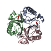
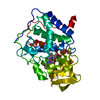
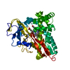
PDB-7in1:
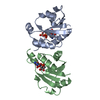
Crystal structure of A2A in complex with TEP for 21h
Method: X-RAY DIFFRACTION / Resolution: 2.49 Å

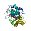
PDB-7in2:
Crystal structure of A2A in complex with 1 mM ZM241385 for 21h
Method: X-RAY DIFFRACTION / Resolution: 2.43 Å

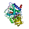
PDB-7in3:
Crystal structure of A2A in complex with 5 mM ZM241385 for 21h
Method: X-RAY DIFFRACTION / Resolution: 2.66 Å

PDB-7in4:
Crystal structure of A2A in complex with 10 mM ZM241385 for 21h
Method: X-RAY DIFFRACTION / Resolution: 2.66 Å

PDB-7in5:
Crystal structure of A2A in complex with TEP for 3h
Method: X-RAY DIFFRACTION / Resolution: 2.49 Å

PDB-7in6:
Crystal structure of A2A in complex with 1 mM ZM241385 for 3h
Method: X-RAY DIFFRACTION / Resolution: 2.52 Å

PDB-7in7:
Crystal structure of A2A in complex with 5 mM ZM241385 for 3h
Method: X-RAY DIFFRACTION / Resolution: 2.43 Å

PDB-7in8:
Crystal structure of A2A in complex with 10 mM ZM241385 for 3h
Method: X-RAY DIFFRACTION / Resolution: 2.52 Å

PDB-7in9:
Crystal structure of A2A in complex with Fi16
Method: X-RAY DIFFRACTION / Resolution: 2.54 Å

PDB-7ina:
Crystal structure of A2A in complex with F2
Method: X-RAY DIFFRACTION / Resolution: 2.15 Å

PDB-7inb:
Crystal structure of A2A in complex with F4
Method: X-RAY DIFFRACTION / Resolution: 2.1 Å

PDB-7inc:
Crystal structure of A2A in complex with F7
Method: X-RAY DIFFRACTION / Resolution: 2.18 Å

PDB-7ind:
Crystal structure of A2A in complex with F12
Method: X-RAY DIFFRACTION / Resolution: 2.67 Å

PDB-7ine:
Crystal structure of A2A in complex with F35
Method: X-RAY DIFFRACTION / Resolution: 2.28 Å

PDB-7inf:
Crystal structure of A2A in complex with F44
Method: X-RAY DIFFRACTION / Resolution: 2.26 Å

PDB-7ing:
Crystal structure of A2A in complex with F99
Method: X-RAY DIFFRACTION / Resolution: 2.15 Å

PDB-7inh:
Crystal structure of A2A in complex with F132
Method: X-RAY DIFFRACTION / Resolution: 2.3 Å

PDB-7ini:
Crystal structure of A2A in complex with F135
Method: X-RAY DIFFRACTION / Resolution: 2.57 Å

PDB-7inj:
Crystal structure of A2A in complex with F148
Method: X-RAY DIFFRACTION / Resolution: 2.1 Å

PDB-7ink:
Crystal structure of A2A in complex with F163
Method: X-RAY DIFFRACTION / Resolution: 2.19 Å

PDB-7inl:
Crystal structure of A2A in complex with F173
Method: X-RAY DIFFRACTION / Resolution: 2.2 Å

PDB-7inm:
Crystal structure of A2A in complex with F178
Method: X-RAY DIFFRACTION / Resolution: 2.19 Å

PDB-7inn:
Crystal structure of A2A in complex with F210
Method: X-RAY DIFFRACTION / Resolution: 2.32 Å

PDB-7ino:
Crystal structure of A2A in complex with F223
Method: X-RAY DIFFRACTION / Resolution: 2.08 Å

PDB-7inp:
Crystal structure of A2A in complex with F235
Method: X-RAY DIFFRACTION / Resolution: 2.21 Å

PDB-7inq:
Crystal structure of A2A in complex with F242
Method: X-RAY DIFFRACTION / Resolution: 2.19 Å

PDB-7inr:
Crystal structure of A2A in complex with F273
Method: X-RAY DIFFRACTION / Resolution: 2.13 Å

PDB-7int:
Crystal structure of A2A in complex with F293
Method: X-RAY DIFFRACTION / Resolution: 2.25 Å

PDB-7inu:
Crystal structure of A2A in complex with F336
Method: X-RAY DIFFRACTION / Resolution: 2.46 Å

PDB-7inv:
Crystal structure of A2A in complex with F379
Method: X-RAY DIFFRACTION / Resolution: 2.57 Å

PDB-7inw:
Crystal structure of A2A in complex with F428
Method: X-RAY DIFFRACTION / Resolution: 2.59 Å

PDB-7inx:
Crystal structure of A2A in complex with FU2-57
Method: X-RAY DIFFRACTION / Resolution: 2.31 Å

PDB-7iny:
Crystal structure of A2A in complex with FU2-59
Method: X-RAY DIFFRACTION / Resolution: 2.36 Å

PDB-7inz:
Crystal structure of A2A in complex with FU4-40
Method: X-RAY DIFFRACTION / Resolution: 2.41 Å

PDB-7io0:
Crystal structure of A2A in complex with FU4-54
Method: X-RAY DIFFRACTION / Resolution: 2.05 Å

PDB-7io1:
Crystal structure of A2A in complex with FU7-1
Method: X-RAY DIFFRACTION / Resolution: 2.25 Å

PDB-7io2:
Crystal structure of A2A in complex with FU7-15
Method: X-RAY DIFFRACTION / Resolution: 1.96 Å

PDB-7io3:
Crystal structure of A2A in complex with FU7-29
Method: X-RAY DIFFRACTION / Resolution: 2.22 Å

PDB-7io4:
Crystal structure of A2A in complex with FU12-19
Method: X-RAY DIFFRACTION / Resolution: 2.1 Å

PDB-7io5:
Crystal structure of A2A in complex with FU12-45
Method: X-RAY DIFFRACTION / Resolution: 1.84 Å

PDB-7io6:
Crystal structure of A2A in complex with FU12-86
Method: X-RAY DIFFRACTION / Resolution: 2.36 Å

PDB-7io7:
Crystal structure of A2A in complex with FU18-60
Method: X-RAY DIFFRACTION / Resolution: 2.23 Å

PDB-7io8:
Crystal structure of A2A in complex with FU18-82
Method: X-RAY DIFFRACTION / Resolution: 2.27 Å

PDB-7io9:
Crystal structure of A2A in complex with FU35-2
Method: X-RAY DIFFRACTION / Resolution: 1.91 Å

PDB-7ioa:
Crystal structure of A2A in complex with FU35-5
Method: X-RAY DIFFRACTION / Resolution: 2.1 Å

PDB-7iob:
Crystal structure of A2A in complex with FU35-42
Method: X-RAY DIFFRACTION / Resolution: 2.11 Å

PDB-7ioc:
Crystal structure of A2A in complex with FU18-82
Method: X-RAY DIFFRACTION / Resolution: 2.07 Å

PDB-7iod:
Crystal structure of A2A in complex with FU35-78
Method: X-RAY DIFFRACTION / Resolution: 2.4 Å

PDB-7ioe:
Crystal structure of A2A in complex with FU44-18
Method: X-RAY DIFFRACTION / Resolution: 2.08 Å

PDB-7iof:
Crystal structure of A2A in complex with FU99-31
Method: X-RAY DIFFRACTION / Resolution: 1.9 Å

PDB-7iog:
Crystal structure of A2A in complex with FU99-89
Method: X-RAY DIFFRACTION / Resolution: 1.98 Å

PDB-7ioh:
Crystal structure of A2A in complex with FU99-91
Method: X-RAY DIFFRACTION / Resolution: 2.07 Å

PDB-7ioi:
Crystal structure of A2A in complex with FU101-88
Method: X-RAY DIFFRACTION / Resolution: 2.04 Å

PDB-7ioj:
Crystal structure of A2A in complex with FU113-13
Method: X-RAY DIFFRACTION / Resolution: 2.26 Å

PDB-7iok:
Crystal structure of A2A in complex with FU113-99
Method: X-RAY DIFFRACTION / Resolution: 2.3 Å

PDB-7iol:
Crystal structure of A2A in complex with FU132-3
Method: X-RAY DIFFRACTION / Resolution: 2.81 Å

PDB-7iom:
Crystal structure of A2A in complex with FU18-82
Method: X-RAY DIFFRACTION / Resolution: 2.1 Å

PDB-7ion:
Crystal structure of A2A in complex with FU132-90
Method: X-RAY DIFFRACTION / Resolution: 2.14 Å

PDB-7ioo:
Crystal structure of A2A in complex with FU132,428-27
Method: X-RAY DIFFRACTION / Resolution: 2.01 Å

PDB-7iop:
Crystal structure of A2A in complex with FU163-33
Method: X-RAY DIFFRACTION / Resolution: 1.92 Å

PDB-7ioq:
Crystal structure of A2A in complex with FU163-51
Method: X-RAY DIFFRACTION / Resolution: 2.26 Å

PDB-7ior:
Crystal structure of A2A in complex with FU163-53
Method: X-RAY DIFFRACTION / Resolution: 1.89 Å

PDB-7ios:
Crystal structure of A2A in complex with FU163-58
Method: X-RAY DIFFRACTION / Resolution: 2.38 Å

PDB-7iot:
Crystal structure of A2A in complex with FU163-67
Method: X-RAY DIFFRACTION / Resolution: 2.31 Å

PDB-7iou:
Crystal structure of A2A in complex with FU163-75
Method: X-RAY DIFFRACTION / Resolution: 2.17 Å

PDB-7iov:
Crystal structure of A2A in complex with FU163-104
Method: X-RAY DIFFRACTION / Resolution: 2.3 Å

PDB-7iow:
Crystal structure of A2A in complex with FU163-106
Method: X-RAY DIFFRACTION / Resolution: 2.17 Å

PDB-7iox:
Crystal structure of A2A in complex with FU163-109
Method: X-RAY DIFFRACTION / Resolution: 2.15 Å

PDB-7ioy:
Crystal structure of A2A in complex with FU210-12
Method: X-RAY DIFFRACTION / Resolution: 2 Å

PDB-7ioz:
Crystal structure of A2A in complex with FU223-7
Method: X-RAY DIFFRACTION / Resolution: 1.88 Å

PDB-7ip0:
Crystal structure of A2A in complex with FU223-43
Method: X-RAY DIFFRACTION / Resolution: 1.99 Å

PDB-7ip1:
Crystal structure of A2A in complex with FU235-66
Method: X-RAY DIFFRACTION / Resolution: 2.29 Å

PDB-7ip2:
Crystal structure of A2A in complex with FU242-98
Method: X-RAY DIFFRACTION / Resolution: 2.13 Å

PDB-7ip3:
Crystal structure of A2A in complex with FU273-23
Method: X-RAY DIFFRACTION / Resolution: 2.11 Å

PDB-7ip4:
Crystal structure of A2A in complex with FU273-77
Method: X-RAY DIFFRACTION / Resolution: 2.04 Å

PDB-7ip5:
Crystal structure of A2A in complex with FU293-69
Method: X-RAY DIFFRACTION / Resolution: 1.95 Å

PDB-7ip6:
Crystal structure of A2A in complex with FU293-80
Method: X-RAY DIFFRACTION / Resolution: 2.07 Å

PDB-7ip7:
Crystal structure of A2A in complex with FU293-87
Method: X-RAY DIFFRACTION / Resolution: 2.32 Å

PDB-7ip8:
Crystal structure of A2A in complex with FU300-24
Method: X-RAY DIFFRACTION / Resolution: 2.28 Å

PDB-7ip9:
Crystal structure of A2A in complex with FU336-22
Method: X-RAY DIFFRACTION / Resolution: 2.1 Å

PDB-7ipa:
Crystal structure of A2A in complex with FU336-32
Method: X-RAY DIFFRACTION / Resolution: 2.32 Å

PDB-7ipb:
Crystal structure of A2A in complex with FU336-34
Method: X-RAY DIFFRACTION / Resolution: 2.09 Å

PDB-7ipc:
Crystal structure of A2A in complex with FU336-61
Method: X-RAY DIFFRACTION / Resolution: 1.87 Å

PDB-7ipd:
Crystal structure of A2A in complex with FU379-101
Method: X-RAY DIFFRACTION / Resolution: 2.19 Å

PDB-7ipe:
Crystal structure of A2A in complex with FU428-8
Method: X-RAY DIFFRACTION / Resolution: 1.84 Å

PDB-7ipf:
Crystal structure of A2A in complex with FU428-16
Method: X-RAY DIFFRACTION / Resolution: 1.9 Å

PDB-7ipg:
Crystal structure of A2A in complex with FU428-36
Method: X-RAY DIFFRACTION / Resolution: 2.06 Å

Chemicals

ChemComp-NA:
Unknown entry

ChemComp-CLR:
CHOLESTEROL

ChemComp-OLA:
OLEIC ACID

ChemComp-OLC:
(2R)-2,3-dihydroxypropyl (9Z)-octadec-9-enoate

ChemComp-HOH:
WATER

PDB-1com:
THE MONOFUNCTIONAL CHORISMATE MUTASE FROM BACILLUS SUBTILIS: STRUCTURE DETERMINATION OF CHORISMATE MUTASE AND ITS COMPLEXES WITH A TRANSITION STATE ANALOG AND PREPHENATE, AND IMPLICATIONS ON THE MECHANISM OF ENZYMATIC REACTION

PDB-1con:
THE REFINED STRUCTURE OF CADMIUM SUBSTITUTED CONCANAVALIN A AT 2.0 ANGSTROMS RESOLUTION

ChemComp-TEP:
THEOPHYLLINE

ChemComp-DMS:
DIMETHYL SULFOXIDE / DMSO, precipitant*YM

ChemComp-8ZX:
4-methoxybenzamide

PDB-1cop:
THREE-DIMENSIONAL DIMER STRUCTURE OF THE LAMBDA-CRO REPRESSOR IN SOLUTION AS DETERMINED BY HETERONUCLEAR MULTIDIMENSIONAL NMR

PDB-1coo:
THE COOH-TERMINAL DOMAIN OF RNA POLYMERASE ALPHA SUBUNIT


ChemComp, No image

ChemComp-VN9:
Unknown entry

ChemComp-ES2:
6-chloro-1,3-dihydro-2H-indol-2-one

PDB-1cor:
INVESTIGATION OF THE SOLUTION CONFORMATION OF CYTOCHROME C-551 FROM PSEUDOMONAS STUTZERI

ChemComp-PEG:
DI(HYDROXYETHYL)ETHER

PDB-1cov:
COXSACKIEVIRUS B3 COAT PROTEIN

PDB-1cow:
BOVINE MITOCHONDRIAL F1-ATPASE COMPLEXED WITH AUROVERTIN B

PDB-1coy:
CRYSTAL STRUCTURE OF CHOLESTEROL OXIDASE COMPLEXED WITH A STEROID SUBSTRATE. IMPLICATIONS FOR FAD DEPENDENT ALCOHOL OXIDASES

ChemComp-U4D:
5-(trifluoromethyl)pyridin-2-one

PDB-1coz:
CTP:GLYCEROL-3-PHOSPHATE CYTIDYLYLTRANSFERASE FROM BACILLUS SUBTILIS

PDB-1co0:
NMR STUDY OF TRP REPRESSOR-MTR OPERATOR DNA COMPLEX

ChemComp-YPN:
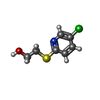
2-[(5-CHLORO-2-PYRIDYL)SULFANYL]ETHANOL

ChemComp-2O8:
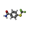
4-[(trifluoromethyl)sulfanyl]benzamide

ChemComp-LR5:
4-(trifluoromethyl)benzamide

PDB-1co1:
FOLD OF THE CBFA

PDB-1co2:
Unknown entry


ChemComp, No image

ChemComp-Y25:
Unknown entry

PDB-1bl1:
PTH RECEPTOR N-TERMINUS FRAGMENT, NMR, 1 STRUCTURE

PDB-1cs6:
N-TERMINAL FRAGMENT OF AXONIN-1 FROM CHICKEN

PDB-1co5:
Unknown entry

PDB-1co6:
CRYSTAL STRUCTURE OF FERROCYTOCHROME C2 FROM RHODOPSEUDOMONAS VIRIDIS

PDB-1co7:
R117H mutant rat anionic trypsin complexed with bovine pancreatic trypsin inhibitor (BPTI)

PDB-1co8:
RECOMBINANT SPERM WHALE MYOGLOBIN L104A MUTANT (MET)

PDB-1co9:
RECOMBINANT SPERM WHALE MYOGLOBIN L104V MUTANT (MET)

PDB-1igl:
SOLUTION STRUCTURE OF HUMAN INSULIN-LIKE GROWTH FACTOR II RELATIONSHIP TO RECEPTOR AND BINDING PROTEIN INTERACTIONS

PDB-1cpb:
STRUCTURE OF CARBOXYPEPTIDASE B AT 2.8 ANGSTROMS RESOLUTION

PDB-1cpc:
ISOLATION, CRYSTALLIZATION, CRYSTAL STRUCTURE ANALYSIS AND REFINEMENT OF CONSTITUTIVE C-PHYCOCYANIN FROM THE CHROMATICALLY ADAPTING CYANOBACTERIUM FREMYELLA DIPLOSIPHON AT 1.66 ANGSTROMS RESOLUTION

PDB-1cpd:
A CATION BINDING MOTIF STABILIZES THE COMPOUND I RADICAL OF CYTOCHROME C PEROXIDASE

PDB-1cpe:
A CATION BINDING MOTIF STABILIZES THE COMPOUND I RADICAL OF CYTOCHROME C PEROXIDASE

PDB-1cpf:
A CATION BINDING MOTIF STABILIZES THE COMPOUND I RADICAL OF CYTOCHROME C PEROXIDASE

PDB-1cph:
CONFORMATIONAL CHANGES IN CUBIC INSULIN CRYSTALS IN THE PH RANGE 7-11

PDB-1cpi:
REGIOSELECTIVE STRUCTURAL AND FUNCTIONAL MIMICRY OF PEPTIDES. DESIGN OF HYDROLYTICALLY STABLE CYCLIC PEPTIDOMIMETIC INHIBITORS OF HIV-1 PROTEASE

PDB-1cpj:
CRYSTAL STRUCTURES OF RECOMBINANT RAT CATHEPSIN B AND A CATHEPSIN B-INHIBITOR COMPLEX: IMPLICATIONS FOR STRUCTURE-BASED INHIBITOR DESIGN

PDB-1cpl:
Unknown entry

PDB-1cpm:
NATIVE-LIKE IN VIVO FOLDING OF A CIRCULARLY PERMUTED JELLYROLL PROTEIN SHOWN BY CRYSTAL STRUCTURE ANALYSIS

PDB-1cpn:
NATIVE-LIKE IN VIVO FOLDING OF A CIRCULARLY PERMUTED JELLYROLL PROTEIN SHOWN BY CRYSTAL STRUCTURE ANALYSIS

PDB-1cpo:
CHLOROPEROXIDASE

PDB-1cpq:
CYTOCHROME C' FROM RHODOPSEUDOMONAS CAPSULATA

PDB-1cpr:
ST. LOUIS CYTOCHROME C' FROM THE PURPLE PHOTOTROPIC BACTERIUM, RHODOBACTER CAPSULATUS

PDB-1cps:
STRUCTURAL COMPARISON OF SULFODIIMINE AND SULFONAMIDE INHIBITORS IN THEIR COMPLEXES WITH ZINC ENZYMES

PDB-1cpt:
CRYSTAL STRUCTURE AND REFINEMENT OF CYTOCHROME P450-TERP AT 2.3 ANGSTROMS RESOLUTION

PDB-1cpu:
SUBSITE MAPPING OF THE ACTIVE SITE OF HUMAN PANCREATIC ALPHA-AMYLASE USING SUBSTRATES, THE PHARMACOLOGICAL INHIBITOR ACARBOSE, AND AN ACTIVE SITE VARIANT

PDB-1cpw:
RECOMBINANT SPERM WHALE MYOGLOBIN L104W MUTANT (MET)

PDB-1cpx:
BETA FORM OF CARBOXYPEPTIDASE A (RESIDUES 3-307) FROM BOVINE PANCREAS IN AN ORTHORHOMBIC CRYSTAL FORM WITH TWO ZINC IONS IN THE ACTIVE SITE.

PDB-1cpy:
SITE-DIRECTED MUTAGENESIS ON (SERINE) CARBOXYPEPTIDASE Y FROM YEAST. THE SIGNIFICANCE OF THR 60 AND MET 398 IN HYDROLYSIS AND AMINOLYSIS REACTIONS

PDB-1cpz:
COPPER CHAPERONE OF ENTEROCOCCUS HIRAE (APO-FORM)

PDB-1cp0:
RECOMBINANT SPERM WHALE MYOGLOBIN L104N MUTANT (MET)

PDB-1cp1:
Unknown entry

PDB-1cp2:
NITROGENASE IRON PROTEIN FROM CLOSTRIDIUM PASTEURIANUM

PDB-1cp3:
CRYSTAL STRUCTURE OF THE COMPLEX OF APOPAIN WITH THE TETRAPEPTIDE INHIBITOR ACE-DVAD-FMC

PDB-1cp4:
FORMATION, CRYSTAL STRUCTURE, AND REARRANGEMENT OF A CYTOCHROME P450-CAM IRON-PHENYL COMPLEX

PDB-1cp5:
RECOMBINANT SPERM WHALE MYOGLOBIN L104F MUTANT (MET)

PDB-1cp6:
1-BUTANEBORONIC ACID BINDING TO AEROMONAS PROTEOLYTICA AMINOPEPTIDASE

PDB-1cp7:
AMINOPEPTIDASE FROM STREPTOMYCES GRISEUS

PDB-1cp8:
NMR STRUCTURE OF DNA (5'-D(TTGGCCAA)2-3') COMPLEXED WITH NOVEL ANTITUMOR DRUG UCH9

PDB-1cp9:
CRYSTAL STRUCTURE OF PENICILLIN G ACYLASE FROM THE BRO1 MUTANT STRAIN OF PROVIDENCIA RETTGERI

PDB-1cqa:
BIRCH POLLEN PROFILIN

ChemComp-6N4:
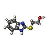
2-(1H-BENZIMIDAZOL-2-YLSULFANYL)ETHANOL

PDB-1cqc:
Unknown entry

PDB-1cqd:
THE 2.1 ANGSTROM STRUCTURE OF A CYSTEINE PROTEASE WITH PROLINE SPECIFICITY FROM GINGER RHIZOME, ZINGIBER OFFICINALE

PDB-1cqe:
PROSTAGLANDIN H2 SYNTHASE-1 COMPLEX WITH FLURBIPROFEN

PDB-1cqf:
THE COMPLEX OF THE MUTATED SHIGA TOXIN B SUBUNIT AND GB3 TRISACCHARIDE

PDB-1cqg:
HIGH RESOLUTION SOLUTION NMR STRUCTURE OF MIXED DISULFIDE INTERMEDIATE BETWEEN HUMAN THIOREDOXIN (C35A, C62A, C69A, C73A) MUTANT AND A 13 RESIDUE PEPTIDE COMPRISING ITS TARGET SITE IN HUMAN REF-1 (RESIDUES 59-71 OF THE P50 SUBUNIT OF NFKB), NMR, 31 STRUCTURES

PDB-1cqh:
HIGH RESOLUTION SOLUTION NMR STRUCTURE OF MIXED DISULFIDE INTERMEDIATE BETWEEN HUMAN THIOREDOXIN (C35A, C62A, C69A, C73A) MUTANT AND A 13 RESIDUE PEPTIDE COMPRISING ITS TARGET SITE IN HUMAN REF-1 (RESIDUES 59-71 OF THE P50 SUBUNIT OF NFKB), NMR, MINIMIZED AVERAGE STRUCTURE

PDB-1cqi:
Crystal Structure of the Complex of ADP and MG2+ with Dephosphorylated E. Coli Succinyl-CoA Synthetase

PDB-1cqj:
CRYSTAL STRUCTURE OF DEPHOSPHORYLATED E. COLI SUCCINYL-COA SYNTHETASE

PDB-1cqk:
CRYSTAL STRUCTURE OF THE CH3 DOMAIN FROM THE MAK33 ANTIBODY

PDB-1cs8:
CRYSTAL STRUCTURE OF PROCATHEPSIN L

PDB-1cql:
NMR STRUCTURE OF SRP RNA DOMAIN IV

Source
  • homo sapiens (human)
  • escherichia coli (E. coli)
KeywordsMEMBRANE PROTEIN / G-PROTEIN-COUPLED RECEPTOR / INTEGRAL MEMBRANE PROTEIN / CHIMERA / 2 THERMOSTABILIZING MUTATIONS

+
About Yorodumi Papers

-
News

-
Feb 9, 2022. New format data for meta-information of EMDB entries

New format data for meta-information of EMDB entries

  • Version 3 of the EMDB header file is now the official format.
  • The previous official version 1.9 will be removed from the archive.

Related info.:EMDB header

External links:wwPDB to switch to version 3 of the EMDB data model

-
Aug 12, 2020. Covid-19 info

Covid-19 info

URL: https://pdbj.org/emnavi/covid19.php

New page: Covid-19 featured information page in EM Navigator.

Related info.:Covid-19 info / Mar 5, 2020. Novel coronavirus structure data

+
Mar 5, 2020. Novel coronavirus structure data

Novel coronavirus structure data

Related info.:Yorodumi Speices / Aug 12, 2020. Covid-19 info

External links:COVID-19 featured content - PDBj / Molecule of the Month (242):Coronavirus Proteases

+
Jan 31, 2019. EMDB accession codes are about to change! (news from PDBe EMDB page)

EMDB accession codes are about to change! (news from PDBe EMDB page)

  • The allocation of 4 digits for EMDB accession codes will soon come to an end. Whilst these codes will remain in use, new EMDB accession codes will include an additional digit and will expand incrementally as the available range of codes is exhausted. The current 4-digit format prefixed with “EMD-” (i.e. EMD-XXXX) will advance to a 5-digit format (i.e. EMD-XXXXX), and so on. It is currently estimated that the 4-digit codes will be depleted around Spring 2019, at which point the 5-digit format will come into force.
  • The EM Navigator/Yorodumi systems omit the EMD- prefix.

Related info.:Q: What is EMD? / ID/Accession-code notation in Yorodumi/EM Navigator

External links:EMDB Accession Codes are Changing Soon! / Contact to PDBj

+
Jul 12, 2017. Major update of PDB

Major update of PDB

  • wwPDB released updated PDB data conforming to the new PDBx/mmCIF dictionary.
  • This is a major update changing the version number from 4 to 5, and with Remediation, in which all the entries are updated.

  • In this update, many items about electron microscopy experimental information are reorganized (e.g. em_software).
  • Now, EM Navigator and Yorodumi are based on the updated data.

External links:wwPDB Remediation / Enriched Model Files Conforming to OneDep Data Standards Now Available in the PDB FTP Archive

-
Yorodumi Papers

Database of articles cited by EMDB/PDB/SASBDB data

  • Database of articles cited by EMDB, PDB, and SASBDB entries
  • Using PubMed data

Related info.:EMDB / PDB / SASBDB / Yorodumi / EMN Papers / Changes in new EM Navigator and Yorodumi

Read more