 Movie
Movie Controller


+ Open data

- Basic information
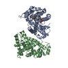
| Entry | Database: PDB / ID: 1cs6 | ||||||
|---|---|---|---|---|---|---|---|
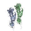
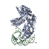
| Title | N-TERMINAL FRAGMENT OF AXONIN-1 FROM CHICKEN | ||||||
 Components Components | AXONIN-1 | ||||||
 Keywords Keywords | CELL ADHESION / NEURAL CELL ADHESION | ||||||
| Function / homology |  Function and homology information Function and homology informationpresynaptic membrane organization / cell-cell adhesion mediator activity / neuron cell-cell adhesion / side of membrane / cell adhesion molecule binding / axon guidance / cell-cell adhesion / brain development / axon / plasma membrane Similarity search - Function | ||||||
| Biological species |  | ||||||
| Method |  X-RAY DIFFRACTION / SYNCHROTRON / Resolution: 1.8 Å X-RAY DIFFRACTION / SYNCHROTRON / Resolution: 1.8 Å | ||||||
 Authors Authors | Freigang, J. / Proba, K. / Diederichs, K. / Sonderegger, P. / Welte, W. | ||||||
 Citation Citation | Journal: Cell(Cambridge,Mass.) / Year: 2000 Title: The crystal structure of the ligand binding module of axonin-1/TAG-1 suggests a zipper mechanism for neural cell adhesion. Authors: Freigang, J. / Proba, K. / Leder, L. / Diederichs, K. / Sonderegger, P. / Welte, W. | ||||||
| History |
|
- Structure visualization
| Structure viewer | Molecule: MolmilJmol/JSmol |
|---|
- Downloads & links
-Download
| PDBx/mmCIF format | 1cs6.cif.gz | 95.4 KB | Display | PDBx/mmCIF format |
|---|---|---|---|---|
| PDB format | pdb1cs6.ent.gz | 71.1 KB | Display | PDB format |
| PDBx/mmJSON format | 1cs6.json.gz | Tree view | PDBx/mmJSON format | |
| Others |  Other downloads Other downloads |
-Validation report
| Arichive directory | https://data.pdbj.org/pub/pdb/validation_reports/cs/1cs6ftp://data.pdbj.org/pub/pdb/validation_reports/cs/1cs6 | HTTPS FTP |
|---|
-Related structure data
| Similar structure data |
|---|


-Links
 PDBj
PDBj


- Assembly
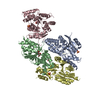
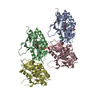
| Deposited unit | 
| ||||||||
|---|---|---|---|---|---|---|---|---|---|
| 1 | 
| ||||||||
| Unit cell |
| ||||||||
| Details | The biological assembly is an oligomer generated by the two-fold screw axis. |
-Components
| #1: Protein | Mass: 42602.734 Da / Num. of mol.: 1 / Fragment: IG1-4 Source method: isolated from a genetically manipulated source Source: (gene. exp.)  |
|---|---|
| #2: Chemical | ChemComp-GOL /   Mass: 92.094 Da / Num. of mol.: 1 / Source method: obtained synthetically / Formula: C3H8O3 Mass: 92.094 Da / Num. of mol.: 1 / Source method: obtained synthetically / Formula: C3H8O3 |
| #3: Water | ChemComp-HOH /  Mass: 18.015 Da / Num. of mol.: 357 / Source method: isolated from a natural source / Formula: H2O Mass: 18.015 Da / Num. of mol.: 357 / Source method: isolated from a natural source / Formula: H2O |
| Has protein modification | Y |
-Experimental details
-Experiment
| Experiment | Method: X-RAY DIFFRACTION / Number of used crystals: 1 |
|---|
- Sample preparation
| Crystal | Density Matthews: 3.2 Å3/Da / Density % sol: 61.6 % | ||||||||||||||||||||||||||||||||||||||||||
|---|---|---|---|---|---|---|---|---|---|---|---|---|---|---|---|---|---|---|---|---|---|---|---|---|---|---|---|---|---|---|---|---|---|---|---|---|---|---|---|---|---|---|---|
| Crystal grow | Temperature: 290 K / Method: vapor diffusion, sitting drop / pH: 7.5 Details: PEG 10000, sodium formiate, HEPES, pH 7.5, VAPOR DIFFUSION, SITTING DROP, temperature 290K | ||||||||||||||||||||||||||||||||||||||||||
| Crystal grow | *PLUS Temperature: 17 ℃ / pH: 8.5 | ||||||||||||||||||||||||||||||||||||||||||
| Components of the solutions | *PLUS
|
-Data collection
| Diffraction | Mean temperature: 100 K |
|---|---|
| Diffraction source | Source: SYNCHROTRON / Site: EMBL/DESY, HAMBURG  / Beamline: X11 / Wavelength: 0.91 / Beamline: X11 / Wavelength: 0.91 |
| Detector | Type: MARRESEARCH / Detector: IMAGE PLATE / Date: Feb 20, 1999 |
| Radiation | Protocol: SINGLE WAVELENGTH / Monochromatic (M) / Laue (L): M / Scattering type: x-ray |
| Radiation wavelength | Wavelength: 0.91 Å / Relative weight: 1 |
| Reflection | Resolution: 1.8→20 Å / Num. all: 50527 / Num. obs: 48051 / % possible obs: 95.1 % / Observed criterion σ(F): 0 / Observed criterion σ(I): 0 / Redundancy: 2.1 % / Biso Wilson estimate: 14.4 Å2 / Rmerge(I) obs: 0.036 / Net I/σ(I): 12.8 |
| Reflection shell | Resolution: 1.8→1.86 Å / Redundancy: 2 % / Rmerge(I) obs: 0.206 / Num. unique all: 3896 / % possible all: 83.3 |
| Reflection shell | *PLUS % possible obs: 83.3 % / Mean I/σ(I) obs: 3.6 |
- Processing
| Software |
| ||||||||||||||||||||||||||||||||||||||||
|---|---|---|---|---|---|---|---|---|---|---|---|---|---|---|---|---|---|---|---|---|---|---|---|---|---|---|---|---|---|---|---|---|---|---|---|---|---|---|---|---|---|
| Refinement | Resolution: 1.8→20 Å / Rfactor Rfree error: 0.005 / Data cutoff high absF: 566597.63 / Data cutoff low absF: 0 / Isotropic thermal model: RESTRAINED / Cross valid method: THROUGHOUT / σ(F): 0 / σ(I): 0 / Stereochemistry target values: Engh & Huber
| ||||||||||||||||||||||||||||||||||||||||
| Solvent computation | Solvent model: FLAT MODEL / Bsol: 48.17 Å2 / ksol: 0.357 e/Å3 | ||||||||||||||||||||||||||||||||||||||||
| Displacement parameters | Biso mean: 33 Å2
| ||||||||||||||||||||||||||||||||||||||||
| Refine analyze |
| ||||||||||||||||||||||||||||||||||||||||
| Refinement step | Cycle: LAST / Resolution: 1.8→20 Å
| ||||||||||||||||||||||||||||||||||||||||
| Refine LS restraints |
| ||||||||||||||||||||||||||||||||||||||||
| LS refinement shell | Resolution: 1.8→1.91 Å / Rfactor Rfree error: 0.016 / Total num. of bins used: 6
| ||||||||||||||||||||||||||||||||||||||||
| Xplor file |
| ||||||||||||||||||||||||||||||||||||||||
| Software | *PLUS Name: CNS / Version: 0.4 / Classification: refinement | ||||||||||||||||||||||||||||||||||||||||
| Refinement | *PLUS σ(F): 0 / % reflection Rfree: 5 % | ||||||||||||||||||||||||||||||||||||||||
| Solvent computation | *PLUS | ||||||||||||||||||||||||||||||||||||||||
| Displacement parameters | *PLUS Biso mean: 33 Å2 | ||||||||||||||||||||||||||||||||||||||||
| Refine LS restraints | *PLUS
| ||||||||||||||||||||||||||||||||||||||||
| LS refinement shell | *PLUS Rfactor Rfree: 0.284 / % reflection Rfree: 4.7 % / Rfactor Rwork: 0.265 |
