 Movie
Movie Controller

[English] 日本語
 Yorodumi
- PDB-4wu3: Structure of the PTP-like myo-inositol phosphatase from Mitsuokel... -
+ Open data

- Basic information
| Entry | Database: PDB / ID: 4wu3 | |||||||||
|---|---|---|---|---|---|---|---|---|---|---|
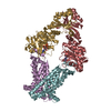
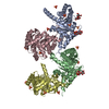
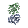
| Title | Structure of the PTP-like myo-inositol phosphatase from Mitsuokella multacida in complex with myo-inositol-(1,3,4,5)-tetrakisphosphate | |||||||||
 Components Components | MYO-INOSITOL PHOSPHOHYDROLASE | |||||||||
 Keywords Keywords | HYDROLASE | |||||||||
| Function / homology |  Function and homology information Function and homology information | |||||||||
| Biological species |  Mitsuokella multacida (bacteria) Mitsuokella multacida (bacteria) | |||||||||
| Method |  X-RAY DIFFRACTION / SYNCHROTRON / MOLECULAR REPLACEMENT / Resolution: 2.2 Å X-RAY DIFFRACTION / SYNCHROTRON / MOLECULAR REPLACEMENT / Resolution: 2.2 Å | |||||||||
 Authors Authors | Bruder, L.M. / Mosimann, S.C. | |||||||||
| Funding support |  Canada, 2items Canada, 2items
| |||||||||
 Citation Citation | Journal: To Be Published Title: Structure of the PTP-like phytase from Selenomonas ruminantium in complex with myo-inositol-(1,3,4,5)-tetrakisphosphate Authors: Bruder, L.M. #1: Journal: J. Mol. Biol. / Year: 2009Title: Structural analysis of a multifunctional, tandemly repeated inositol polyphosphatase. Authors: Gruninger, R.J. / Selinger, L.B. / Mosimann, S.C. | |||||||||
| History |
|
- Structure visualization
| Structure viewer | Molecule: MolmilJmol/JSmol |
|---|
- Downloads & links
-Download
| PDBx/mmCIF format | 4wu3.cif.gz | 535.4 KB | Display | PDBx/mmCIF format |
|---|---|---|---|---|
| PDB format | pdb4wu3.ent.gz | 436.5 KB | Display | PDB format |
| PDBx/mmJSON format | 4wu3.json.gz | Tree view | PDBx/mmJSON format | |
| Others |  Other downloads Other downloads |
-Validation report
| Arichive directory | https://data.pdbj.org/pub/pdb/validation_reports/wu/4wu3ftp://data.pdbj.org/pub/pdb/validation_reports/wu/4wu3 | HTTPS FTP |
|---|
-Related structure data
| Related structure data |  4wtyC  3f41S S: Starting model for refinement C: citing same article ( |
|---|---|
| Similar structure data |


-Links
 PDBj
PDBj


- Assembly
| Deposited unit | 
| ||||||||||||||||||||||||||||||||||||||||||||||||||||||||||||||||||||||||||||||||||||||||||||||||||
|---|---|---|---|---|---|---|---|---|---|---|---|---|---|---|---|---|---|---|---|---|---|---|---|---|---|---|---|---|---|---|---|---|---|---|---|---|---|---|---|---|---|---|---|---|---|---|---|---|---|---|---|---|---|---|---|---|---|---|---|---|---|---|---|---|---|---|---|---|---|---|---|---|---|---|---|---|---|---|---|---|---|---|---|---|---|---|---|---|---|---|---|---|---|---|---|---|---|---|---|
| 1 | 
| ||||||||||||||||||||||||||||||||||||||||||||||||||||||||||||||||||||||||||||||||||||||||||||||||||
| 2 | 
| ||||||||||||||||||||||||||||||||||||||||||||||||||||||||||||||||||||||||||||||||||||||||||||||||||
| Unit cell |
| ||||||||||||||||||||||||||||||||||||||||||||||||||||||||||||||||||||||||||||||||||||||||||||||||||
| Noncrystallographic symmetry (NCS) | NCS domain:
NCS domain segments: Component-ID: 1 / Beg auth comp-ID: PRO / Beg label comp-ID: PRO / End auth comp-ID: ALA / End label comp-ID: ALA / Refine code: 1 / Auth seq-ID: 47 - 636 / Label seq-ID: 39 - 628
NCS ensembles :
|
-Components
| #1: Protein | Mass: 72448.023 Da / Num. of mol.: 4 / Fragment: UNP residues 32-640 / Mutation: C250S, C548S Source method: isolated from a genetically manipulated source Details: Inactive with C252S/C548S mutations / Source: (gene. exp.) Mitsuokella multacida (bacteria) / Gene: phyA / Plasmid: pET28b / Production host: #2: Chemical | ChemComp-4IP /   Mass: 500.075 Da / Num. of mol.: 6 / Source method: obtained synthetically / Formula: C6H16O18P4 Mass: 500.075 Da / Num. of mol.: 6 / Source method: obtained synthetically / Formula: C6H16O18P4#3: Chemical | ChemComp-PO4 /   Mass: 94.971 Da / Num. of mol.: 6 / Source method: obtained synthetically / Formula: PO4 Mass: 94.971 Da / Num. of mol.: 6 / Source method: obtained synthetically / Formula: PO4#4: Chemical | ChemComp-GOL /   Mass: 92.094 Da / Num. of mol.: 12 / Source method: obtained synthetically / Formula: C3H8O3 Mass: 92.094 Da / Num. of mol.: 12 / Source method: obtained synthetically / Formula: C3H8O3#5: Water | ChemComp-HOH / |  Mass: 18.015 Da / Num. of mol.: 2262 / Source method: isolated from a natural source / Formula: H2O Mass: 18.015 Da / Num. of mol.: 2262 / Source method: isolated from a natural source / Formula: H2O |
|---|
-Experimental details
-Experiment
| Experiment | Method: X-RAY DIFFRACTION / Number of used crystals: 1 |
|---|
- Sample preparation
| Crystal | Density Matthews: 2.62 Å3/Da / Density % sol: 53.04 % |
|---|---|
| Crystal grow | Temperature: 293 K / Method: vapor diffusion, sitting drop / pH: 8 Details: PEG 8000, Polyethylene glycol, Tris chloride, beta-mercapto ethanol, glycerol |
-Data collection
| Diffraction | Mean temperature: 100 K |
|---|---|
| Diffraction source | Source: SYNCHROTRON / Site: CLSI / Beamline: 08ID-1 / Wavelength: 0.97934 Å |
| Detector | Type: RAYONIX MX-300 / Detector: CCD / Date: Feb 4, 2011 |
| Radiation | Protocol: SINGLE WAVELENGTH / Monochromatic (M) / Laue (L): M / Scattering type: x-ray |
| Radiation wavelength | Wavelength: 0.97934 Å / Relative weight: 1 |
| Reflection | Resolution: 2.2→44.54 Å / Num. obs: 146418 / % possible obs: 98.8 % / Redundancy: 2.2 % / Rmerge(I) obs: 0.08 / Net I/σ(I): 6 |
| Reflection shell | Resolution: 2.2→2.24 Å / Redundancy: 2.2 % / Rmerge(I) obs: 0.16 / Mean I/σ(I) obs: 4.3 / % possible all: 96.6 |
- Processing
| Software |
| |||||||||||||||||||||||||||||||||||||||||||||||||||||||||||||||||||||||||||
|---|---|---|---|---|---|---|---|---|---|---|---|---|---|---|---|---|---|---|---|---|---|---|---|---|---|---|---|---|---|---|---|---|---|---|---|---|---|---|---|---|---|---|---|---|---|---|---|---|---|---|---|---|---|---|---|---|---|---|---|---|---|---|---|---|---|---|---|---|---|---|---|---|---|---|---|---|
| Refinement | Method to determine structure: MOLECULAR REPLACEMENT Starting model: 3F41 Resolution: 2.2→44.54 Å / Cor.coef. Fo:Fc: 0.923 / Cor.coef. Fo:Fc free: 0.906 / SU B: 6.052 / SU ML: 0.148 / Cross valid method: THROUGHOUT / ESU R: 0.26 / ESU R Free: 0.184 / Stereochemistry target values: MAXIMUM LIKELIHOOD / Details: HYDROGENS HAVE BEEN ADDED IN THE RIDING POSITIONS
| |||||||||||||||||||||||||||||||||||||||||||||||||||||||||||||||||||||||||||
| Solvent computation | Ion probe radii: 0.8 Å / Shrinkage radii: 0.8 Å / VDW probe radii: 1.2 Å / Solvent model: MASK | |||||||||||||||||||||||||||||||||||||||||||||||||||||||||||||||||||||||||||
| Displacement parameters | Biso mean: 19.523 Å2
| |||||||||||||||||||||||||||||||||||||||||||||||||||||||||||||||||||||||||||
| Refinement step | Cycle: LAST / Resolution: 2.2→44.54 Å
| |||||||||||||||||||||||||||||||||||||||||||||||||||||||||||||||||||||||||||
| Refine LS restraints |
| |||||||||||||||||||||||||||||||||||||||||||||||||||||||||||||||||||||||||||
| Refine LS restraints NCS | Dom-ID: 1 / Number: 5556 / Refine-ID: X-RAY DIFFRACTION / Type: TIGHT THERMAL / Weight position: 0.87
| |||||||||||||||||||||||||||||||||||||||||||||||||||||||||||||||||||||||||||
| LS refinement shell | Resolution: 2.2→2.257 Å / Total num. of bins used: 20
|
