 Movie
Movie Controller

[English] 日本語
 Yorodumi
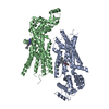
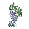
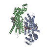
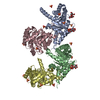
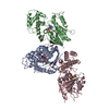
- PDB-1xcf: Crystal structure of P28L/Y173F tryptophan synthase alpha-subunit... -
+ Open data

- Basic information
| Entry | Database: PDB / ID: 1xcf | ||||||
|---|---|---|---|---|---|---|---|
| Title | Crystal structure of P28L/Y173F tryptophan synthase alpha-subunits from Escherichia coli | ||||||
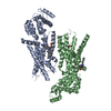
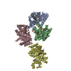
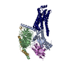
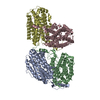
 Components Components | Tryptophan synthase alpha chain | ||||||
 Keywords Keywords | LYASE / tryptophan synthase / a-subunits / E.coli / P28L/Y173F double mutants | ||||||
| Function / homology |  Function and homology information Function and homology informationtryptophan synthase / tryptophan synthase activity / L-tryptophan biosynthetic process / aromatic amino acid biosynthetic process / molecular adaptor activity / lyase activity / cytoplasm / cytosol Similarity search - Function | ||||||
| Biological species |  | ||||||
| Method |  X-RAY DIFFRACTION / SYNCHROTRON / MOLECULAR REPLACEMENT / Resolution: 1.8 Å X-RAY DIFFRACTION / SYNCHROTRON / MOLECULAR REPLACEMENT / Resolution: 1.8 Å | ||||||
 Authors Authors | Jang, S.B. | ||||||
 Citation Citation | Journal: Biochem.Biophys.Res.Commun. / Year: 2004 Title: Structures of wild-type and P28L/Y173F tryptophan synthase alpha-subunits from Escherichia coli Authors: Jeong, M.S. / Jeong, J.K. / Lim, W.K. / Jang, S.B. #1: Journal: J.Mol.Biol. / Year: 2002Title: On the role of alphaThr183 in the allosteric regulation and catalytic mechanism of tryptophan synthase Authors: Kulik, V. / Weyand, M. / Seidel, R. / Niks, D. / Arac, D. / Dunn, M.F. / Schlichting, I. | ||||||
| History |
|
- Structure visualization
| Structure viewer | Molecule: MolmilJmol/JSmol |
|---|
- Downloads & links
-Download
| PDBx/mmCIF format | 1xcf.cif.gz | 105.6 KB | Display | PDBx/mmCIF format |
|---|---|---|---|---|
| PDB format | pdb1xcf.ent.gz | 81.2 KB | Display | PDB format |
| PDBx/mmJSON format | 1xcf.json.gz | Tree view | PDBx/mmJSON format | |
| Others |  Other downloads Other downloads |
-Validation report
| Arichive directory | https://data.pdbj.org/pub/pdb/validation_reports/xc/1xcfftp://data.pdbj.org/pub/pdb/validation_reports/xc/1xcf | HTTPS FTP |
|---|
-Related structure data
| Related structure data | 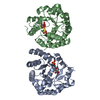 1xc4C  1k8xS S: Starting model for refinement C: citing same article ( |
|---|---|
| Similar structure data |

-Links
 PDBj
PDBj
- Assembly
| Deposited unit | 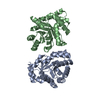
| ||||||||
|---|---|---|---|---|---|---|---|---|---|
| 1 | 
| ||||||||
| Unit cell |
|
-Components
| #1: Protein | Mass: 28782.188 Da / Num. of mol.: 2 / Mutation: P28L/Y173F Source method: isolated from a genetically manipulated source Source: (gene. exp.) #2: Water | ChemComp-HOH / |  Mass: 18.015 Da / Num. of mol.: 231 / Source method: isolated from a natural source / Formula: H2O Mass: 18.015 Da / Num. of mol.: 231 / Source method: isolated from a natural source / Formula: H2O |
|---|
-Experimental details
-Experiment
| Experiment | Method: X-RAY DIFFRACTION / Number of used crystals: 1 |
|---|
- Sample preparation
| Crystal | Density Matthews: 2.34 Å3/Da / Density % sol: 47.52 % |
|---|---|
| Crystal grow | Temperature: 298 K / Method: vapor diffusion, hanging drop / pH: 7.5 Details: HEPES-NA, isopropanol, polyethylene glycol4000, pH 7.5, VAPOR DIFFUSION, HANGING DROP, temperature 298K |
-Data collection
| Diffraction | Mean temperature: 100 K |
|---|---|
| Diffraction source | Source: SYNCHROTRON / Site: PAL/PLS  / Beamline: 6B / Wavelength: 1.12714 Å / Beamline: 6B / Wavelength: 1.12714 Å |
| Detector | Type: MACSCIENCE / Detector: IMAGE PLATE / Date: Nov 26, 2003 |
| Radiation | Monochromator: copper / Protocol: SINGLE WAVELENGTH / Monochromatic (M) / Laue (L): M / Scattering type: x-ray |
| Radiation wavelength | Wavelength: 1.12714 Å / Relative weight: 1 |
| Reflection | Resolution: 1.8→30 Å / Num. all: 387319 / Num. obs: 48396 / % possible obs: 91.9 % / Observed criterion σ(F): 1 / Observed criterion σ(I): 1 / Redundancy: 8 % / Biso Wilson estimate: 29.4 Å2 / Rmerge(I) obs: 0.054 / Net I/σ(I): 24.8 |
| Reflection shell | Resolution: 1.8→1.86 Å / Rmerge(I) obs: 0.289 / Num. unique all: 3485 / % possible all: 79.7 |
- Processing
| Software |
| |||||||||||||||||||||||||
|---|---|---|---|---|---|---|---|---|---|---|---|---|---|---|---|---|---|---|---|---|---|---|---|---|---|---|
| Refinement | Method to determine structure: MOLECULAR REPLACEMENT Starting model: 1K8X Resolution: 1.8→30 Å / σ(F): 1 / Stereochemistry target values: Engh & Huber
| |||||||||||||||||||||||||
| Displacement parameters | Biso mean: 29.4 Å2 | |||||||||||||||||||||||||
| Refinement step | Cycle: LAST / Resolution: 1.8→30 Å
| |||||||||||||||||||||||||
| Refine LS restraints |
| |||||||||||||||||||||||||
| LS refinement shell | Resolution: 1.8→1.86 Å
|
