 Movie
Movie Controller

[English] 日本語
 Yorodumi
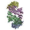
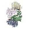
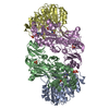
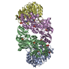
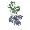
- PDB-1cqi: Crystal Structure of the Complex of ADP and MG2+ with Dephosphory... -
+ Open data

- Basic information
| Entry | Database: PDB / ID: 1cqi | ||||||
|---|---|---|---|---|---|---|---|
| Title | Crystal Structure of the Complex of ADP and MG2+ with Dephosphorylated E. Coli Succinyl-CoA Synthetase | ||||||
 Components Components | (PROTEIN (SUCCINYL-COA SYNTHETASE ...) x 2 | ||||||
 Keywords Keywords | LIGASE / ATP-GRASP FOLD / ROSSMANN FOLD | ||||||
| Function / homology |  Function and homology information Function and homology informationsuccinate-CoA ligase (GDP-forming) activity / succinate-CoA ligase complex (ADP-forming) / succinate-CoA ligase (ADP-forming) / succinate-CoA ligase complex / succinate-CoA ligase (ADP-forming) activity / succinyl-CoA metabolic process / tricarboxylic acid cycle / nucleotide binding / magnesium ion binding / ATP binding ...succinate-CoA ligase (GDP-forming) activity / succinate-CoA ligase complex (ADP-forming) / succinate-CoA ligase (ADP-forming) / succinate-CoA ligase complex / succinate-CoA ligase (ADP-forming) activity / succinyl-CoA metabolic process / tricarboxylic acid cycle / nucleotide binding / magnesium ion binding / ATP binding / cytoplasm / cytosol Similarity search - Function | ||||||
| Biological species |  | ||||||
| Method |  X-RAY DIFFRACTION / SYNCHROTRON / Resolution: 3.3 Å X-RAY DIFFRACTION / SYNCHROTRON / Resolution: 3.3 Å | ||||||
 Authors Authors | Joyce, M.A. / Fraser, M.E. / James, M.N.G. / Bridger, W.A. / Wolodko, W.T. | ||||||
 Citation Citation | Journal: Biochemistry / Year: 2000 Title: ADP-binding site of Escherichia coli succinyl-CoA synthetase revealed by x-ray crystallography. Authors: Joyce, M.A. / Fraser, M.E. / James, M.N. / Bridger, W.A. / Wolodko, W.T. #1: Journal: J.Mol.Biol. / Year: 1999Title: A Detailed Structural Description of Escherichia Coli Succinyl-Coa Synthetase Authors: Fraser, M.E. / James, M.N.G. / Bridger, W.A. / Wolodko, W.T. #2: Journal: J.Biol.Chem. / Year: 1994Title: The Crystal Structure of Succinyl-Coa Synthetase from Escherichia Coli at 2.5A Resolution Authors: Wolodko, W.T. / Fraser, M.E. / James, M.N.G. / Bridger, W.A. #3: Journal: J.Biol.Chem. / Year: 1984Title: Crystallization of Succinyl-Coa Synthetase from Escherichia Coli Authors: Wolodko, W.T. / James, M.N.G. / Bridger, W.A. | ||||||
| History |
|
- Structure visualization
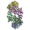
| Structure viewer | Molecule: MolmilJmol/JSmol |
|---|
- Downloads & links
-Download
| PDBx/mmCIF format | 1cqi.cif.gz | 237.2 KB | Display | PDBx/mmCIF format |
|---|---|---|---|---|
| PDB format | pdb1cqi.ent.gz | 193.5 KB | Display | PDB format |
| PDBx/mmJSON format | 1cqi.json.gz | Tree view | PDBx/mmJSON format | |
| Others |  Other downloads Other downloads |
-Validation report
| Arichive directory | https://data.pdbj.org/pub/pdb/validation_reports/cq/1cqiftp://data.pdbj.org/pub/pdb/validation_reports/cq/1cqi | HTTPS FTP |
|---|
-Related structure data

-Links
 PDBj
PDBj


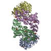
- Assembly
| Deposited unit | 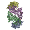
| ||||||||||
|---|---|---|---|---|---|---|---|---|---|---|---|
| 1 |
| ||||||||||
| Unit cell |
|
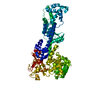
-Components
-PROTEIN (SUCCINYL-COA SYNTHETASE ... , 2 types, 4 molecules ADBE
| #1: Protein | Mass: 29436.902 Da / Num. of mol.: 2 / Fragment: ALPHA SUBUNIT Source method: isolated from a genetically manipulated source Source: (gene. exp.) References: UniProt: P07459, UniProt: P0AGE9*PLUS, succinate-CoA ligase (ADP-forming) #2: Protein | Mass: 41123.152 Da / Num. of mol.: 2 / Fragment: BETA SUBUNIT Source method: isolated from a genetically manipulated source Source: (gene. exp.) References: UniProt: P07460, UniProt: P0A836*PLUS, succinate-CoA ligase (ADP-forming) |
|---|
-Non-polymers , 4 types, 8 molecules 


| #3: Chemical |  Mass: 94.971 Da / Num. of mol.: 2 / Source method: obtained synthetically / Formula: PO4 Mass: 94.971 Da / Num. of mol.: 2 / Source method: obtained synthetically / Formula: PO4#4: Chemical |  Mass: 767.534 Da / Num. of mol.: 2 / Source method: obtained synthetically / Formula: C21H36N7O16P3S Mass: 767.534 Da / Num. of mol.: 2 / Source method: obtained synthetically / Formula: C21H36N7O16P3S#5: Chemical |  Mass: 24.305 Da / Num. of mol.: 2 / Source method: obtained synthetically / Formula: Mg Mass: 24.305 Da / Num. of mol.: 2 / Source method: obtained synthetically / Formula: Mg#6: Chemical |  Mass: 427.201 Da / Num. of mol.: 2 / Source method: obtained synthetically / Formula: C10H15N5O10P2 / Comment: ADP, energy-carrying molecule*YM Mass: 427.201 Da / Num. of mol.: 2 / Source method: obtained synthetically / Formula: C10H15N5O10P2 / Comment: ADP, energy-carrying molecule*YM |
|---|
-Details
| Has protein modification | Y |
|---|
-Experimental details
-Experiment
| Experiment | Method: X-RAY DIFFRACTION / Number of used crystals: 6 |
|---|
- Sample preparation
| Crystal | Density Matthews: 3.59 Å3/Da / Density % sol: 65.78 % | ||||||||||||||||||||||||||||||
|---|---|---|---|---|---|---|---|---|---|---|---|---|---|---|---|---|---|---|---|---|---|---|---|---|---|---|---|---|---|---|---|
| Crystal grow | Method: vapor diffusion, hanging drop / pH: 7.3 Details: AMMONIUM SULFATE, POTASSIUM PHOSPHATE, COENZYME A, pH 7.30, VAPOR DIFFUSION, HANGING DROP | ||||||||||||||||||||||||||||||
| Crystal grow | *PLUS Temperature: 21 ℃ / Method: microdialysis | ||||||||||||||||||||||||||||||
| Components of the solutions | *PLUS
|
-Data collection
| Diffraction | Mean temperature: 283 K |
|---|---|
| Diffraction source | Source: SYNCHROTRON / Site: Photon Factory  / Beamline: BL-6A / Wavelength: 1 / Beamline: BL-6A / Wavelength: 1 |
| Detector | Type: FUJI / Detector: IMAGE PLATE / Date: Jun 8, 1996 |
| Radiation | Protocol: SINGLE WAVELENGTH / Monochromatic (M) / Laue (L): M / Scattering type: x-ray |
| Radiation wavelength | Wavelength: 1 Å / Relative weight: 1 |
| Reflection | Resolution: 3.3→20 Å / Num. all: 31753 / Num. obs: 31753 / % possible obs: 99 % / Observed criterion σ(I): 0 / Redundancy: 6.3 % / Biso Wilson estimate: 22.8 Å2 / Rmerge(I) obs: 0.164 |
| Reflection shell | Resolution: 3.3→3.5 Å / Redundancy: 3.5 % / Rmerge(I) obs: 0.378 / % possible all: 98.4 |
| Reflection | *PLUS Num. measured all: 195264 |
| Reflection shell | *PLUS Lowest resolution: 3.54 Å / Num. unique obs: 15389 |
- Processing
| Software |
| ||||||||||||||||||||||||||||||||||||||||||||||||||||||||||||
|---|---|---|---|---|---|---|---|---|---|---|---|---|---|---|---|---|---|---|---|---|---|---|---|---|---|---|---|---|---|---|---|---|---|---|---|---|---|---|---|---|---|---|---|---|---|---|---|---|---|---|---|---|---|---|---|---|---|---|---|---|---|
| Refinement | Resolution: 3.3→20 Å / Rfactor Rfree error: 0.004 / Isotropic thermal model: GROUP / Cross valid method: THROUGHOUT / σ(F): 0 / Stereochemistry target values: ENGH & HUBER / Details: MAXIMUM LIKELIHOOD REFINEMENT IN CNS
| ||||||||||||||||||||||||||||||||||||||||||||||||||||||||||||
| Solvent computation | Solvent model: FLAT MODEL / Bsol: 10 Å2 / ksol: 0.217 e/Å3 | ||||||||||||||||||||||||||||||||||||||||||||||||||||||||||||
| Displacement parameters | Biso mean: 34.7 Å2 | ||||||||||||||||||||||||||||||||||||||||||||||||||||||||||||
| Refine analyze |
| ||||||||||||||||||||||||||||||||||||||||||||||||||||||||||||
| Refinement step | Cycle: LAST / Resolution: 3.3→20 Å
| ||||||||||||||||||||||||||||||||||||||||||||||||||||||||||||
| Refine LS restraints |
| ||||||||||||||||||||||||||||||||||||||||||||||||||||||||||||
| Refine LS restraints NCS | NCS model details: CONSTR | ||||||||||||||||||||||||||||||||||||||||||||||||||||||||||||
| LS refinement shell | Resolution: 3.3→3.51 Å / Rfactor Rfree error: 0.014 / Total num. of bins used: 6
| ||||||||||||||||||||||||||||||||||||||||||||||||||||||||||||
| Software | *PLUS Name: 'CNS' / Classification: refinement | ||||||||||||||||||||||||||||||||||||||||||||||||||||||||||||
| Refine LS restraints | *PLUS
|
