 Movie
Movie Controller

[English] 日本語
 Yorodumi
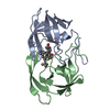
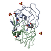
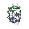
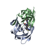
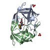
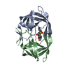
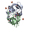
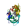
- PDB-1cpi: REGIOSELECTIVE STRUCTURAL AND FUNCTIONAL MIMICRY OF PEPTIDES. DES... -
+ Open data

- Basic information
| Entry | Database: PDB / ID: 1cpi | ||||||
|---|---|---|---|---|---|---|---|
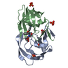
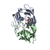
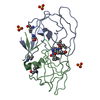
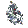
| Title | REGIOSELECTIVE STRUCTURAL AND FUNCTIONAL MIMICRY OF PEPTIDES. DESIGN OF HYDROLYTICALLY STABLE CYCLIC PEPTIDOMIMETIC INHIBITORS OF HIV-1 PROTEASE | ||||||
 Components Components |
| ||||||
 Keywords Keywords | HYDROLASE/HYDROLASE INHIBITOR / HYDROLASE-HYDROLASE INHIBITOR complex | ||||||
| Function / homology |  Function and homology information Function and homology informationHIV-1 retropepsin / symbiont-mediated activation of host apoptosis / retroviral ribonuclease H / exoribonuclease H / exoribonuclease H activity / DNA integration / viral genome integration into host DNA / establishment of integrated proviral latency / RNA-directed DNA polymerase / RNA stem-loop binding ...HIV-1 retropepsin / symbiont-mediated activation of host apoptosis / retroviral ribonuclease H / exoribonuclease H / exoribonuclease H activity / DNA integration / viral genome integration into host DNA / establishment of integrated proviral latency / RNA-directed DNA polymerase / RNA stem-loop binding / viral penetration into host nucleus / host multivesicular body / RNA-directed DNA polymerase activity / RNA-DNA hybrid ribonuclease activity / Transferases; Transferring phosphorus-containing groups; Nucleotidyltransferases / host cell / viral nucleocapsid / DNA recombination / DNA-directed DNA polymerase / aspartic-type endopeptidase activity / Hydrolases; Acting on ester bonds / DNA-directed DNA polymerase activity / symbiont-mediated suppression of host gene expression / viral translational frameshifting / symbiont entry into host cell / lipid binding / host cell nucleus / host cell plasma membrane / virion membrane / structural molecule activity / proteolysis / DNA binding / zinc ion binding Similarity search - Function | ||||||
| Method |  X-RAY DIFFRACTION / Resolution: 2.05 Å X-RAY DIFFRACTION / Resolution: 2.05 Å | ||||||
 Authors Authors | Martin, J.L. | ||||||
 Citation Citation | Journal: J.Am.Chem.Soc. / Year: 1995 Title: REGIOSELECTIVE STRUCTURAL AND FUNCTIONAL MIMICRY OF PEPTIDES - DESIGN OF HYDROLYTICALLY-STABLE CYCLIC PEPTIDOMIMETIC INHIBITORS OF HIV-1 PROTEASE. Authors: Abbenante, G. / March, D.R. / Bergman, D.A. / Hunt, P.A. / Garnham, B. / Dancer, R.J. / Martin, J.L. / Fairlie, D.P. | ||||||
| History |
|
- Structure visualization
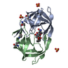
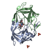
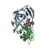
| Structure viewer | Molecule: MolmilJmol/JSmol |
|---|
- Downloads & links
-Download
| PDBx/mmCIF format | 1cpi.cif.gz | 53.8 KB | Display | PDBx/mmCIF format |
|---|---|---|---|---|
| PDB format | pdb1cpi.ent.gz | 38.3 KB | Display | PDB format |
| PDBx/mmJSON format | 1cpi.json.gz | Tree view | PDBx/mmJSON format | |
| Others |  Other downloads Other downloads |
-Validation report
| Arichive directory | https://data.pdbj.org/pub/pdb/validation_reports/cp/1cpiftp://data.pdbj.org/pub/pdb/validation_reports/cp/1cpi | HTTPS FTP |
|---|
-Related structure data
| Similar structure data |
|---|

-Links
 PDBj
PDBj

- Assembly
| Deposited unit | 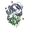
| ||||||||
|---|---|---|---|---|---|---|---|---|---|
| 1 |
| ||||||||
| Unit cell |
|
-Components
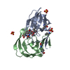
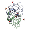
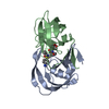
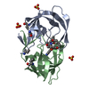
| #1: Protein | Mass: 10764.636 Da / Num. of mol.: 2 / Source method: obtained synthetically Details: CHEMICALLY SYNTHESIZED PROTEIN CORRESPONDING TO THE PROTEASE FROM THE HIV-I References: UniProt: P03369 #2: Protein/peptide | |   Type: Peptide-like / Class: Inhibitor / Mass: 767.731 Da / Num. of mol.: 1 / Source method: obtained synthetically Type: Peptide-like / Class: Inhibitor / Mass: 767.731 Da / Num. of mol.: 1 / Source method: obtained syntheticallyReferences: 1-{(2R)-2-[(8R,11S)-8-(2-amino-2-oxoethyl)-6,9-dioxo-2-oxa-7,10-diazabicyclo[11.2.2]heptadeca-1(15),13,16-trien-11-yl]- 2-hydroxyethyl}-L-prolyl-L-isoleucyl-L-valinamide #3: Chemical |   Mass: 96.063 Da / Num. of mol.: 3 / Source method: obtained synthetically / Formula: SO4 Mass: 96.063 Da / Num. of mol.: 3 / Source method: obtained synthetically / Formula: SO4#4: Water | ChemComp-HOH / |  Mass: 18.015 Da / Num. of mol.: 95 / Source method: isolated from a natural source / Formula: H2O Mass: 18.015 Da / Num. of mol.: 95 / Source method: isolated from a natural source / Formula: H2O |
|---|
-Experimental details
-Experiment
| Experiment | Method: X-RAY DIFFRACTION |
|---|
- Sample preparation
| Crystal | Density Matthews: 2.12 Å3/Da / Density % sol: 41.94 % | |||||||||||||||
|---|---|---|---|---|---|---|---|---|---|---|---|---|---|---|---|---|
| Crystal grow | pH: 5.5 / Details: pH 5.5 | |||||||||||||||
| Crystal | *PLUS Density % sol: 44 % | |||||||||||||||
| Crystal grow | *PLUS Temperature: 20 ℃ / Method: vapor diffusion, hanging drop | |||||||||||||||
| Components of the solutions | *PLUS
|
-Data collection
| Diffraction | Mean temperature: 289 K |
|---|---|
| Diffraction source | Wavelength: 1.5418 Å |
| Detector | Type: RIGAKU / Detector: IMAGE PLATE / Date: Dec 10, 1993 |
| Radiation | Monochromator: GRAPHITE / Monochromatic (M) / Laue (L): M / Scattering type: x-ray |
| Radiation wavelength | Wavelength: 1.5418 Å / Relative weight: 1 |
| Reflection | Resolution: 2.05→51 Å / Num. obs: 11539 / % possible obs: 94 % / Observed criterion σ(I): 1 / Redundancy: 3 % / Rmerge(I) obs: 0.071 |
| Reflection | *PLUS Rmerge(I) obs: 0.071 |
- Processing
| Software |
| ||||||||||||||||||||||||||||||||||||||||||||||||||||||||||||
|---|---|---|---|---|---|---|---|---|---|---|---|---|---|---|---|---|---|---|---|---|---|---|---|---|---|---|---|---|---|---|---|---|---|---|---|---|---|---|---|---|---|---|---|---|---|---|---|---|---|---|---|---|---|---|---|---|---|---|---|---|---|
| Refinement | Resolution: 2.05→8 Å / σ(F): 2
| ||||||||||||||||||||||||||||||||||||||||||||||||||||||||||||
| Displacement parameters | Biso mean: 20.9 Å2 | ||||||||||||||||||||||||||||||||||||||||||||||||||||||||||||
| Refine analyze | Luzzati coordinate error obs: 0.2 Å | ||||||||||||||||||||||||||||||||||||||||||||||||||||||||||||
| Refinement step | Cycle: LAST / Resolution: 2.05→8 Å
| ||||||||||||||||||||||||||||||||||||||||||||||||||||||||||||
| Refine LS restraints |
| ||||||||||||||||||||||||||||||||||||||||||||||||||||||||||||
| Software | *PLUS Name: X-PLOR / Classification: refinement | ||||||||||||||||||||||||||||||||||||||||||||||||||||||||||||
| Refinement | *PLUS | ||||||||||||||||||||||||||||||||||||||||||||||||||||||||||||
| Solvent computation | *PLUS | ||||||||||||||||||||||||||||||||||||||||||||||||||||||||||||
| Displacement parameters | *PLUS | ||||||||||||||||||||||||||||||||||||||||||||||||||||||||||||
| Refine LS restraints | *PLUS
|
