 Movie
Movie Controller

[English] 日本語
 Yorodumi
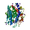
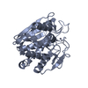
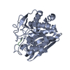
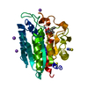
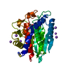
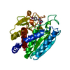
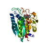
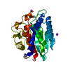
- PDB-1cp6: 1-BUTANEBORONIC ACID BINDING TO AEROMONAS PROTEOLYTICA AMINOPEPTIDASE -
+ Open data

- Basic information
| Entry | Database: PDB / ID: 1cp6 | |||||||||
|---|---|---|---|---|---|---|---|---|---|---|
| Title | 1-BUTANEBORONIC ACID BINDING TO AEROMONAS PROTEOLYTICA AMINOPEPTIDASE | |||||||||
 Components Components | PROTEIN (AMINOPEPTIDASE) | |||||||||
 Keywords Keywords | HYDROLASE / AMINOPEPTIDASE | |||||||||
| Function / homology |  Function and homology information Function and homology informationbacterial leucyl aminopeptidase / metalloexopeptidase activity / aminopeptidase activity / proteolysis / extracellular region / metal ion binding Similarity search - Function | |||||||||
| Biological species |  Vibrio proteolyticus (bacteria) Vibrio proteolyticus (bacteria) | |||||||||
| Method |  X-RAY DIFFRACTION / OTHER / Resolution: 1.9 Å X-RAY DIFFRACTION / OTHER / Resolution: 1.9 Å | |||||||||
 Authors Authors | Depaola, C.C. / Bennett, B. / Holz, R.C. / Ringe, D. / Petsko, G.A. | |||||||||
 Citation Citation | Journal: Biochemistry / Year: 1999 Title: 1-Butaneboronic acid binding to Aeromonas proteolytica aminopeptidase: a case of arrested development. Authors: De Paola, C.C. / Bennett, B. / Holz, R.C. / Ringe, D. / Petsko, G.A. #1: Journal: Structure / Year: 1994Title: Crystal Structure of the Aeromonas Proteolytica Aminopeptidase: A Prototypical Member of the Co-Catalytic Zinc Family Authors: Chevrier, B. / Schalk, C. / D'Orchmont, H. / Rondeau, J.-M. / Moras, D. / Tarnus, C. | |||||||||
| History |
|
- Structure visualization
| Structure viewer | Molecule: MolmilJmol/JSmol |
|---|
- Downloads & links
-Download
| PDBx/mmCIF format | 1cp6.cif.gz | 70.6 KB | Display | PDBx/mmCIF format |
|---|---|---|---|---|
| PDB format | pdb1cp6.ent.gz | 52 KB | Display | PDB format |
| PDBx/mmJSON format | 1cp6.json.gz | Tree view | PDBx/mmJSON format | |
| Others |  Other downloads Other downloads |
-Validation report
| Arichive directory | https://data.pdbj.org/pub/pdb/validation_reports/cp/1cp6ftp://data.pdbj.org/pub/pdb/validation_reports/cp/1cp6 | HTTPS FTP |
|---|
-Related structure data
| Related structure data |  1ampS S: Starting model for refinement |
|---|---|
| Similar structure data |


-Links
 PDBj
PDBj
- Assembly
| Deposited unit | 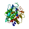
| |||||||||
|---|---|---|---|---|---|---|---|---|---|---|
| 1 |
| |||||||||
| Unit cell |
| |||||||||
| Components on special symmetry positions |
|
-Components
| #1: Protein | Mass: 31427.350 Da / Num. of mol.: 1 / Source method: isolated from a natural source / Source: (natural) Vibrio proteolyticus (bacteria)References: UniProt: Q01693, bacterial leucyl aminopeptidase | ||||||
|---|---|---|---|---|---|---|---|
| #2: Chemical |   Mass: 65.409 Da / Num. of mol.: 2 / Source method: obtained synthetically / Formula: Zn Mass: 65.409 Da / Num. of mol.: 2 / Source method: obtained synthetically / Formula: Zn#3: Chemical | ChemComp-BUB / |   Mass: 101.940 Da / Num. of mol.: 1 / Source method: obtained synthetically / Formula: C4H11BO2 Mass: 101.940 Da / Num. of mol.: 1 / Source method: obtained synthetically / Formula: C4H11BO2#4: Water | ChemComp-HOH / |  Mass: 18.015 Da / Num. of mol.: 136 / Source method: isolated from a natural source / Formula: H2O Mass: 18.015 Da / Num. of mol.: 136 / Source method: isolated from a natural source / Formula: H2OHas protein modification | Y | |
-Experimental details
-Experiment
| Experiment | Method: X-RAY DIFFRACTION / Number of used crystals: 1 |
|---|
- Sample preparation
| Crystal | Density Matthews: 2.67 Å3/Da / Density % sol: 54.01 % | |||||||||||||||||||||||||||||||||||||||||||||||||||||||||||||||
|---|---|---|---|---|---|---|---|---|---|---|---|---|---|---|---|---|---|---|---|---|---|---|---|---|---|---|---|---|---|---|---|---|---|---|---|---|---|---|---|---|---|---|---|---|---|---|---|---|---|---|---|---|---|---|---|---|---|---|---|---|---|---|---|---|
| Crystal grow | pH: 8 / Details: 100 MM TRIS PH 8.0, 100 MM KSCN, 4.5 M NACL | |||||||||||||||||||||||||||||||||||||||||||||||||||||||||||||||
| Crystal grow | *PLUS Method: vapor diffusion | |||||||||||||||||||||||||||||||||||||||||||||||||||||||||||||||
| Components of the solutions | *PLUS
|
-Data collection
| Diffraction | Mean temperature: 277 K |
|---|---|
| Diffraction source | Source: ROTATING ANODE / Type: RIGAKU RU200 / Wavelength: 1.5418 |
| Detector | Type: RIGAKU RAXIS / Detector: IMAGE PLATE / Date: Mar 15, 1997 |
| Radiation | Monochromator: NI FILTER / Protocol: SINGLE WAVELENGTH / Monochromatic (M) / Laue (L): M / Scattering type: x-ray |
| Radiation wavelength | Wavelength: 1.5418 Å / Relative weight: 1 |
| Reflection | Resolution: 1.9→30 Å / Num. obs: 23658 / % possible obs: 84.2 % / Observed criterion σ(I): 0 / Redundancy: 6.9 % / Rmerge(I) obs: 0.134 |
| Reflection shell | Resolution: 1.9→2.1 Å / Rmerge(I) obs: 0.378 / % possible all: 46.7 |
| Reflection | *PLUS Num. measured all: 163530 |
| Reflection shell | *PLUS % possible obs: 46.7 % |
- Processing
| Software |
| ||||||||||||||||||||||||||||||||||||||||||||||||||||||||||||
|---|---|---|---|---|---|---|---|---|---|---|---|---|---|---|---|---|---|---|---|---|---|---|---|---|---|---|---|---|---|---|---|---|---|---|---|---|---|---|---|---|---|---|---|---|---|---|---|---|---|---|---|---|---|---|---|---|---|---|---|---|---|
| Refinement | Method to determine structure: OTHER Starting model: 1AMP Resolution: 1.9→10 Å / Data cutoff high absF: 0 / Data cutoff low absF: 0 / Cross valid method: THROUGHOUT / σ(F): 0
| ||||||||||||||||||||||||||||||||||||||||||||||||||||||||||||
| Refinement step | Cycle: LAST / Resolution: 1.9→10 Å
| ||||||||||||||||||||||||||||||||||||||||||||||||||||||||||||
| Refine LS restraints |
| ||||||||||||||||||||||||||||||||||||||||||||||||||||||||||||
| LS refinement shell | Resolution: 1.9→10 Å / Total num. of bins used: 8 / % reflection obs: 85 % | ||||||||||||||||||||||||||||||||||||||||||||||||||||||||||||
| Software | *PLUS Name: X-PLOR / Version: 4 / Classification: refinement | ||||||||||||||||||||||||||||||||||||||||||||||||||||||||||||
| Refine LS restraints | *PLUS
|
