 Movie
Movie Controller

[English] 日本語
 Yorodumi
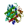
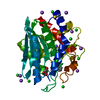
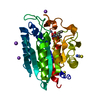
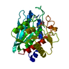
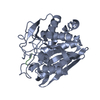
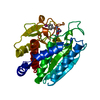
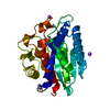
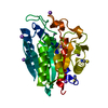
- PDB-2nyq: Structure of Vibrio proteolyticus aminopeptidase with a bound Trp... -
+ Open data

- Basic information
| Entry | Database: PDB / ID: 2nyq | ||||||
|---|---|---|---|---|---|---|---|
| Title | Structure of Vibrio proteolyticus aminopeptidase with a bound Trp fragment of dLWCF | ||||||
 Components Components |
| ||||||
 Keywords Keywords | HYDROLASE / Trp / VpAP / non-covalent | ||||||
| Function / homology |  Function and homology information Function and homology informationbacterial leucyl aminopeptidase / metalloexopeptidase activity / aminopeptidase activity / proteolysis / extracellular region / metal ion binding Similarity search - Function | ||||||
| Biological species |  Vibrio proteolyticus (bacteria) Vibrio proteolyticus (bacteria)synthetic construct (others) | ||||||
| Method |  X-RAY DIFFRACTION / MOLECULAR REPLACEMENT / Resolution: 2.5 Å X-RAY DIFFRACTION / MOLECULAR REPLACEMENT / Resolution: 2.5 Å | ||||||
 Authors Authors | Bennett, B. / Kumar, A. / Narayanan, B. / Kim, J.-J. | ||||||
 Citation Citation | Journal: To be Published Title: Substrate recognition by the leucine aminopeptidase from Vibrio proteolyticus Authors: Kumar, A. / Narayanan, B. / Kim, J.-J. / Bennett, B. | ||||||
| History |
|
- Structure visualization
| Structure viewer | Molecule: MolmilJmol/JSmol |
|---|
- Downloads & links
-Download
| PDBx/mmCIF format | 2nyq.cif.gz | 71.3 KB | Display | PDBx/mmCIF format |
|---|---|---|---|---|
| PDB format | pdb2nyq.ent.gz | 53.3 KB | Display | PDB format |
| PDBx/mmJSON format | 2nyq.json.gz | Tree view | PDBx/mmJSON format | |
| Others |  Other downloads Other downloads |
-Validation report
| Arichive directory | https://data.pdbj.org/pub/pdb/validation_reports/ny/2nyqftp://data.pdbj.org/pub/pdb/validation_reports/ny/2nyq | HTTPS FTP |
|---|
-Related structure data
| Related structure data |  1cp6S S: Starting model for refinement |
|---|---|
| Similar structure data |


-Links
 PDBj
PDBj
- Assembly
| Deposited unit | 
| ||||||||
|---|---|---|---|---|---|---|---|---|---|
| 1 |
| ||||||||
| Unit cell |
|
-Components
| #1: Protein | Mass: 32238.150 Da / Num. of mol.: 1 / Fragment: Bacterial leucyl aminopeptidase, residues 97-405 / Source method: isolated from a natural source / Source: (natural) Vibrio proteolyticus (bacteria) / Strain: ATTC 15338, DSM 30189, NCBM 1326References: UniProt: Q01693, bacterial leucyl aminopeptidase | ||||
|---|---|---|---|---|---|
| #2: Protein/peptide | Mass: 567.699 Da / Num. of mol.: 1 / Source method: obtained synthetically Details: The tetrapeptide was chemically synthesised using solid-phase manual synthesis via Fmoc-derivatized amino acids, on Rink-amide resin. Source: (synth.) synthetic construct (others) | ||||
| #3: Chemical |   Mass: 65.409 Da / Num. of mol.: 2 / Source method: obtained synthetically / Formula: Zn Mass: 65.409 Da / Num. of mol.: 2 / Source method: obtained synthetically / Formula: Zn#4: Water | ChemComp-HOH / |  Mass: 18.015 Da / Num. of mol.: 152 / Source method: isolated from a natural source / Formula: H2O Mass: 18.015 Da / Num. of mol.: 152 / Source method: isolated from a natural source / Formula: H2OHas protein modification | Y | |
-Experimental details
-Experiment
| Experiment | Method: X-RAY DIFFRACTION / Number of used crystals: 1 |
|---|
- Sample preparation
| Crystal | Density Matthews: 2.55 Å3/Da / Density % sol: 51.67 % |
|---|---|
| Crystal grow | Temperature: 296 K / Method: vapor diffusion, sitting drop / pH: 8 Details: 5 d., 10/100/100 mM KSCN, 0.4/4.5/4.5 M NaCl, 10/100 mM Tris/100 mM Tricine, pH 8.0, VAPOR DIFFUSION, SITTING DROP, temperature 296K |
-Data collection
| Diffraction | Mean temperature: 100 K |
|---|---|
| Diffraction source | Source: ROTATING ANODE / Type: RIGAKU / Wavelength: 1.54 Å |
| Detector | Type: RIGAKU RAXIS IV / Detector: IMAGE PLATE / Date: Jun 1, 2005 / Details: CONFOCAL |
| Radiation | Monochromator: MIRROR / Protocol: SINGLE WAVELENGTH / Monochromatic (M) / Laue (L): M / Scattering type: x-ray |
| Radiation wavelength | Wavelength: 1.54 Å / Relative weight: 1 |
| Reflection | Resolution: 2.5→50 Å / Num. all: 12122 / Num. obs: 12122 / % possible obs: 98.4 % / Biso Wilson estimate: 36.5 Å2 / Rsym value: 0.048 |
| Reflection shell | Resolution: 2.5→2.59 Å / Rsym value: 0.139 / % possible all: 97.7 |
- Processing
| Software |
| ||||||||||||||||||||
|---|---|---|---|---|---|---|---|---|---|---|---|---|---|---|---|---|---|---|---|---|---|
| Refinement | Method to determine structure: MOLECULAR REPLACEMENT Starting model: PDB entry 1CP6 Resolution: 2.5→50 Å
| ||||||||||||||||||||
| Refinement step | Cycle: LAST / Resolution: 2.5→50 Å
|
