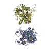
PDB-3sd7: 1.7 Angstrom Resolution Crystal Structure of Putative Phosphatase from Clostridium difficile 手法: X-RAY DIFFRACTION / 解像度: 1.7 Å
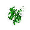
PDB-3srt: The crystal structure of a maltose O-acetyltransferase from Clostridium difficile 630 手法: X-RAY DIFFRACTION / 解像度: 2.504 Å
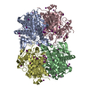
PDB-3uuw: 1.63 Angstrom Resolution Crystal Structure of Dehydrogenase (MviM) from Clostridium difficile. 手法: X-RAY DIFFRACTION / 解像度: 1.63 Å
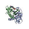
PDB-4dd5: Biosynthetic Thiolase (ThlA1) from Clostridium difficile 手法: X-RAY DIFFRACTION / 解像度: 1.25 Å
PDB-4dgt: Crystal structure of PLP-bound putative aminotransferase from Clostridium difficile 630 crystallized with magnesium formate 手法: X-RAY DIFFRACTION / 解像度: 1.55 Å
PDB-4dq6: Crystal structure of PLP-bound putative aminotransferase from Clostridium difficile 630 手法: X-RAY DIFFRACTION / 解像度: 1.5 Å
PDB-4dun: 1.76A X-ray Crystal Structure of a Putative Phenazine Biosynthesis PhzC/PhzF Protein from Clostridium difficile (strain 630) 手法: X-RAY DIFFRACTION / 解像度: 1.76 Å
PDB-4e1l: Crystal structure of Acetoacetyl-CoA thiolase (thlA2) from Clostridium difficile 手法: X-RAY DIFFRACTION / 解像度: 2 Å
PDB-4egu: 0.95A Resolution Structure of a Histidine Triad Protein from Clostridium difficile 手法: X-RAY DIFFRACTION / 解像度: 0.95 Å
PDB-4gib: 2.27 Angstrom Crystal Structure of beta-Phosphoglucomutase (pgmB) from Clostridium difficile 手法: X-RAY DIFFRACTION / 解像度: 2.27 Å
PDB-4h3d: 1.95 Angstrom Crystal Structure of of Type I 3-Dehydroquinate Dehydratase (aroD) from Clostridium difficile with Covalent Modified Comenic Acid. 手法: X-RAY DIFFRACTION / 解像度: 1.95 Å
PDB-4isx: The crystal structure of maltose o-acetyltransferase from clostridium difficile 630 in complex with acetyl-coa 手法: X-RAY DIFFRACTION / 解像度: 2.702 Å
PDB-4jjp: 2.06 Angstrom resolution crystal structure of phosphomethylpyrimidine kinase (thiD)from Clostridium difficile 630 手法: X-RAY DIFFRACTION / 解像度: 2.056 Å
PDB-4kd5: substrate binding domain of putative molybdenum ABC transporter from Clostridium difficile 手法: X-RAY DIFFRACTION / 解像度: 2.4999 Å
PDB-4mfg: 2.0 Angstrom Resolution Crystal Structure of Putative Carbonic Anhydrase from Clostridium difficile. 手法: X-RAY DIFFRACTION / 解像度: 2 Å
PDB-4nmy: Crystal Structure of the Thiamin-bound form of Substrate-binding Protein of ABC Transporter from Clostridium difficile 手法: X-RAY DIFFRACTION / 解像度: 1.896 Å
PDB-4rn7: The crystal structure of N-acetylmuramoyl-L-alanine amidase from Clostridium difficile 630 手法: X-RAY DIFFRACTION / 解像度: 1.717 Å
PDB-5dzs: 1.5 Angstrom Crystal Structure of Shikimate Dehydrogenase 1 from Peptoclostridium difficile. 手法: X-RAY DIFFRACTION / 解像度: 1.5 Å
PDB-5tta: A 1.85A X-Ray Structure from Peptoclostridium difficile 630 of a Hypothetical Protein 手法: X-RAY DIFFRACTION / 解像度: 1.85 Å
PDB-5tv7: 2.05 Angstrom Resolution Crystal Structure of Peptidoglycan-Binding Protein from Clostridioides difficile in Complex with Glutamine Hydroxamate. 手法: X-RAY DIFFRACTION / 解像度: 2.05 Å
PDB-5txu: 1.95 Angstrom Resolution Crystal Structure of Stage II Sporulation Protein D (SpoIID) from Clostridium difficile in Apo Conformation 手法: X-RAY DIFFRACTION / 解像度: 1.95 Å
PDB-6n7m: 1.78 Angstrom Resolution Crystal Structure of Hypothetical Protein CD630_05490 from Clostridioides difficile 630. 手法: X-RAY DIFFRACTION / 解像度: 1.78 Å
PDB-6ue2: 1.85 Angstrom Resolution Crystal Structure of Class D beta-lactamase from Clostridium difficile 630 手法: X-RAY DIFFRACTION / 解像度: 1.85 Å
PDB-6wy4: Crystal Structure of Wild Type Class D beta-lactamase from Clostridium difficile 630 手法: X-RAY DIFFRACTION / 解像度: 1.8 Å
PDB-7k1u: Crystal Structure of SrtB-anchored Collagen-binding Adhesin Fragment (residues 206-565) from Clostridioides difficile strain 630 手法: X-RAY DIFFRACTION / 解像度: 2.4 Å
PDB-7rl8: Crystal Structure of C79A Mutant of Class D beta-lactamase from Clostridium difficile 630 手法: X-RAY DIFFRACTION / 解像度: 1.95 Å
PDB-7rlr: Crystal Structure of K83A Mutant of Class D beta-lactamase from Clostridium difficile 630 手法: X-RAY DIFFRACTION / 解像度: 1.88 Å
 ムービー
ムービー コントローラー
 構造ビューア
 万見文献について


 著者
著者 リンク
リンク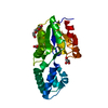


 キーワード
キーワード clostridium difficile (バクテリア)