 Movie
Movie Controller

[English] 日本語
 Yorodumi
- PDB-6n7m: 1.78 Angstrom Resolution Crystal Structure of Hypothetical Protei... -
+ Open data

- Basic information
| Entry | Database: PDB / ID: 6n7m | ||||||
|---|---|---|---|---|---|---|---|
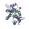
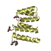
| Title | 1.78 Angstrom Resolution Crystal Structure of Hypothetical Protein CD630_05490 from Clostridioides difficile 630. | ||||||
 Components Components | Hypothetical Protein CD630_05490 | ||||||
 Keywords Keywords | UNKNOWN FUNCTION / Structural Genomics / Center for Structural Genomics of Infectious Diseases / CSGID / Hypothetical Protein | ||||||
| Function / homology | Uncharacterized protein Function and homology information Function and homology information | ||||||
| Biological species |  Peptoclostridium difficile (bacteria) Peptoclostridium difficile (bacteria) | ||||||
| Method |  X-RAY DIFFRACTION / SYNCHROTRON / SAD / Resolution: 1.78 Å X-RAY DIFFRACTION / SYNCHROTRON / SAD / Resolution: 1.78 Å | ||||||
 Authors Authors | Minasov, G. / Shuvalova, L. / Wawrzak, Z. / Kiryukhina, O. / Dubrovska, I. / Anderson, W.F. / Satchell, K.J.F. / Joachimiak, A. / Center for Structural Genomics of Infectious Diseases (CSGID) | ||||||
 Citation Citation | Journal: Microbiol Resour Announc / Year: 2023 Title: A high-throughput structural system biology approach to increase structure representation of proteins from Clostridioides difficile. Authors: Rosas-Lemus, M. / Dey, S. / Minasov, G. / Tan, K. / Anderson, S.M. / Brunzelle, J. / Nocadello, S. / Shabalin, I. / Filippova, E. / Halavaty, A. / Kim, Y. / Maltseva, N. / Osipiuk, J. / ...Authors: Rosas-Lemus, M. / Dey, S. / Minasov, G. / Tan, K. / Anderson, S.M. / Brunzelle, J. / Nocadello, S. / Shabalin, I. / Filippova, E. / Halavaty, A. / Kim, Y. / Maltseva, N. / Osipiuk, J. / Minor, W. / Joachimiak, A. / Savchenko, A. / Anderson, W.F. / Satchell, K.J.F. | ||||||
| History |
|
- Structure visualization
| Structure viewer | Molecule: MolmilJmol/JSmol |
|---|
- Downloads & links
-Download
| PDBx/mmCIF format | 6n7m.cif.gz | 75.5 KB | Display | PDBx/mmCIF format |
|---|---|---|---|---|
| PDB format | pdb6n7m.ent.gz | 56 KB | Display | PDB format |
| PDBx/mmJSON format | 6n7m.json.gz | Tree view | PDBx/mmJSON format | |
| Others |  Other downloads Other downloads |
-Validation report
| Arichive directory | https://data.pdbj.org/pub/pdb/validation_reports/n7/6n7mftp://data.pdbj.org/pub/pdb/validation_reports/n7/6n7m | HTTPS FTP |
|---|
-Related structure data
| Related structure data | 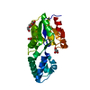 3sd7C  3srtC  3uuwC 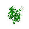 4dd5C  4dgtC  4dq6C  4dunC 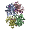 4e1lC 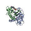 4eguC  4gibC  4h3dC  4isxC  4jjpC  4kd5C  4mfgC  4nmyC  4rn7C  5dzsC  5ttaC  5tv7C  5txuC  6ue2C  6wy4C  7k1uC  7rl8C  7rlrC C: citing same article ( |
|---|---|
| Similar structure data | |
| Other databases |


-Links
 PDBj
PDBj- Assembly
| Deposited unit | 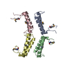
| ||||||||||||||||||||||||||||||||||||||||||||||||||||||||||||||||||||||||||||||||||||||||||||||||||||||||||||||||||||||||||||||||||||||||||||||||||||||
|---|---|---|---|---|---|---|---|---|---|---|---|---|---|---|---|---|---|---|---|---|---|---|---|---|---|---|---|---|---|---|---|---|---|---|---|---|---|---|---|---|---|---|---|---|---|---|---|---|---|---|---|---|---|---|---|---|---|---|---|---|---|---|---|---|---|---|---|---|---|---|---|---|---|---|---|---|---|---|---|---|---|---|---|---|---|---|---|---|---|---|---|---|---|---|---|---|---|---|---|---|---|---|---|---|---|---|---|---|---|---|---|---|---|---|---|---|---|---|---|---|---|---|---|---|---|---|---|---|---|---|---|---|---|---|---|---|---|---|---|---|---|---|---|---|---|---|---|---|---|---|---|
| 1 | 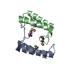
| ||||||||||||||||||||||||||||||||||||||||||||||||||||||||||||||||||||||||||||||||||||||||||||||||||||||||||||||||||||||||||||||||||||||||||||||||||||||
| 2 | 
| ||||||||||||||||||||||||||||||||||||||||||||||||||||||||||||||||||||||||||||||||||||||||||||||||||||||||||||||||||||||||||||||||||||||||||||||||||||||
| Unit cell |
| ||||||||||||||||||||||||||||||||||||||||||||||||||||||||||||||||||||||||||||||||||||||||||||||||||||||||||||||||||||||||||||||||||||||||||||||||||||||
| Noncrystallographic symmetry (NCS) | NCS domain:
NCS domain segments: Component-ID: _ / Beg auth comp-ID: GLY / Beg label comp-ID: GLY / Refine code: _
NCS ensembles :
|
-Components
| #1: Protein | Mass: 12624.758 Da / Num. of mol.: 4 Source method: isolated from a genetically manipulated source Source: (gene. exp.) Peptoclostridium difficile (strain 630) (bacteria)Strain: 630 / Gene: CD630_05490 / Plasmid: pMCSG53 / Production host: #2: Water | ChemComp-HOH / |  Mass: 18.015 Da / Num. of mol.: 213 / Source method: isolated from a natural source / Formula: H2O Mass: 18.015 Da / Num. of mol.: 213 / Source method: isolated from a natural source / Formula: H2OHas protein modification | Y | |
|---|
-Experimental details
-Experiment
| Experiment | Method: X-RAY DIFFRACTION / Number of used crystals: 1 |
|---|
- Sample preparation
| Crystal | Density Matthews: 1.19 Å3/Da / Density % sol: 39.38 % |
|---|---|
| Crystal grow | Temperature: 292 K / Method: vapor diffusion, sitting drop / pH: 8.3 Details: Protein: 9.3 mg/ml, 0.01M Tris HCl (pH 8.3); Screen: Classics II (H11), 0.1M Potassium thiocyanate, 30% (w/v) PEG 2000 MME. |
-Data collection
| Diffraction | Mean temperature: 100 K / Serial crystal experiment: N |
|---|---|
| Diffraction source | Source: SYNCHROTRON / Site: APS  / Beamline: 21-ID-F / Wavelength: 0.97872 Å / Beamline: 21-ID-F / Wavelength: 0.97872 Å |
| Detector | Type: MARMOSAIC 300 mm CCD / Detector: CCD / Date: Apr 20, 2017 / Details: C(111) |
| Radiation | Monochromator: Be / Protocol: SINGLE WAVELENGTH / Monochromatic (M) / Laue (L): M / Scattering type: x-ray |
| Radiation wavelength | Wavelength: 0.97872 Å / Relative weight: 1 |
| Reflection | Resolution: 1.78→46.7 Å / Num. obs: 22345 / % possible obs: 97.74 % / Redundancy: 5.2 % / CC1/2: 0.99 / Rmerge(I) obs: 0.102 / Rpim(I) all: 0.048 / Rrim(I) all: 0.113 / Rsym value: 0.102 / Net I/σ(I): 11.1 |
| Reflection shell | Resolution: 1.78→1.81 Å / Redundancy: 4.1 % / Rmerge(I) obs: 0.83 / Mean I/σ(I) obs: 2.1 / Num. unique obs: 1068 / CC1/2: 0.61 / Rpim(I) all: 0.45 / Rrim(I) all: 0.95 / Rsym value: 0.83 / % possible all: 97.89 |
- Processing
| Software |
| ||||||||||||||||||||||||||||||||||||||||||||||||||||||||||||||||||||||||||||||||||||||||||||||||||||||||||||||||||||||||||||||||||||||||||||||||||||||||||||||||||||||||||||||||||||||
|---|---|---|---|---|---|---|---|---|---|---|---|---|---|---|---|---|---|---|---|---|---|---|---|---|---|---|---|---|---|---|---|---|---|---|---|---|---|---|---|---|---|---|---|---|---|---|---|---|---|---|---|---|---|---|---|---|---|---|---|---|---|---|---|---|---|---|---|---|---|---|---|---|---|---|---|---|---|---|---|---|---|---|---|---|---|---|---|---|---|---|---|---|---|---|---|---|---|---|---|---|---|---|---|---|---|---|---|---|---|---|---|---|---|---|---|---|---|---|---|---|---|---|---|---|---|---|---|---|---|---|---|---|---|---|---|---|---|---|---|---|---|---|---|---|---|---|---|---|---|---|---|---|---|---|---|---|---|---|---|---|---|---|---|---|---|---|---|---|---|---|---|---|---|---|---|---|---|---|---|---|---|---|---|
| Refinement | Method to determine structure: SAD / Resolution: 1.78→24.52 Å / Cor.coef. Fo:Fc: 0.919 / Cor.coef. Fo:Fc free: 0.898 / SU B: 5.118 / SU ML: 0.157 / Cross valid method: THROUGHOUT / ESU R: 0.211 / ESU R Free: 0.177 / Details: HYDROGENS HAVE BEEN ADDED IN THE RIDING POSITIONS
| ||||||||||||||||||||||||||||||||||||||||||||||||||||||||||||||||||||||||||||||||||||||||||||||||||||||||||||||||||||||||||||||||||||||||||||||||||||||||||||||||||||||||||||||||||||||
| Solvent computation | Ion probe radii: 0.8 Å / Shrinkage radii: 0.8 Å / VDW probe radii: 1.2 Å | ||||||||||||||||||||||||||||||||||||||||||||||||||||||||||||||||||||||||||||||||||||||||||||||||||||||||||||||||||||||||||||||||||||||||||||||||||||||||||||||||||||||||||||||||||||||
| Displacement parameters | Biso mean: 29.866 Å2
| ||||||||||||||||||||||||||||||||||||||||||||||||||||||||||||||||||||||||||||||||||||||||||||||||||||||||||||||||||||||||||||||||||||||||||||||||||||||||||||||||||||||||||||||||||||||
| Refinement step | Cycle: 1 / Resolution: 1.78→24.52 Å
| ||||||||||||||||||||||||||||||||||||||||||||||||||||||||||||||||||||||||||||||||||||||||||||||||||||||||||||||||||||||||||||||||||||||||||||||||||||||||||||||||||||||||||||||||||||||
| Refine LS restraints |
|