 Movie
Movie Controller


+ Open data

- Basic information
| Entry | Database: PDB / ID: 5ibo | ||||||
|---|---|---|---|---|---|---|---|
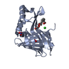
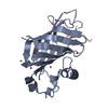
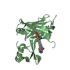
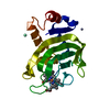
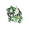
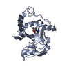
| Title | 1.95A resolution structure of NanoLuc luciferase | ||||||
 Components Components | Oplophorus-luciferin 2-monooxygenase catalytic subunit | ||||||
 Keywords Keywords | OXIDOREDUCTASE / Oplophorus bioluminescent protein / NanoLuc luciferase / NLuc / coelenterazine / furimazine / beta-barrel | ||||||
| Function / homology | Oplophorus-luciferin 2-monooxygenase / Oplophorus-luciferin 2-monooxygenase activity / Calycin / bioluminescence / extracellular region / DECANOIC ACID / Oplophorus-luciferin 2-monooxygenase catalytic subunit Function and homology information Function and homology information | ||||||
| Biological species |  Oplophorus gracilirostris (arthropod) Oplophorus gracilirostris (arthropod) | ||||||
| Method |  X-RAY DIFFRACTION / SYNCHROTRON / MOLECULAR REPLACEMENT / molecular replacement / Resolution: 1.95 Å X-RAY DIFFRACTION / SYNCHROTRON / MOLECULAR REPLACEMENT / molecular replacement / Resolution: 1.95 Å | ||||||
 Authors Authors | Lovell, S. / Mehzabeen, N. / Battaile, K.P. / Wood, M.G. / Encell, L.P. / Wood, K.V. | ||||||
 Citation Citation | Journal: To be published Title: To be determined Authors: Lovell, S. / Mehzabeen, N. / Battaile, K.P. / Wood, M.G. / Encell, L.P. / Wood, K.V. | ||||||
| History |
|
- Structure visualization
| Structure viewer | Molecule: MolmilJmol/JSmol |
|---|
- Downloads & links
-Download
| PDBx/mmCIF format | 5ibo.cif.gz | 142.9 KB | Display | PDBx/mmCIF format |
|---|---|---|---|---|
| PDB format | pdb5ibo.ent.gz | 112.8 KB | Display | PDB format |
| PDBx/mmJSON format | 5ibo.json.gz | Tree view | PDBx/mmJSON format | |
| Others |  Other downloads Other downloads |
-Validation report
| Arichive directory | https://data.pdbj.org/pub/pdb/validation_reports/ib/5iboftp://data.pdbj.org/pub/pdb/validation_reports/ib/5ibo | HTTPS FTP |
|---|
-Related structure data
| Related structure data | 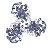 4dyaC 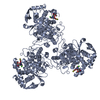 4dybC 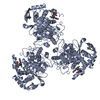 4dynC 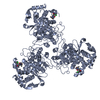 4dypC  4dysC  4dytC  6dqcC  6dqdC  6dqeC  6dqfC  7rt0C  9bqnC C: citing same article ( |
|---|---|
| Similar structure data |


-Links
 PDBj
PDBj

- Assembly
| Deposited unit | 
| ||||||||
|---|---|---|---|---|---|---|---|---|---|
| 1 | 
| ||||||||
| 2 | 
| ||||||||
| Unit cell |
|
-Components
| #1: Protein | Mass: 19429.164 Da / Num. of mol.: 2 / Fragment: Full Length Mutation: A4E, Q11R, Q18L, L27V, A33N, K43R, V44I, A54I, F68D, L72Q, M75K, I90V, P115E, Q124K, Y138I, N166R Source method: isolated from a genetically manipulated source Source: (gene. exp.) Oplophorus gracilirostris (arthropod) / Plasmid: pFN18K(-AIA) / Production host:  References: UniProt: Q9GV45, Oplophorus-luciferin 2-monooxygenase #2: Chemical |   Mass: 172.265 Da / Num. of mol.: 2 / Source method: obtained synthetically / Formula: C10H20O2 Mass: 172.265 Da / Num. of mol.: 2 / Source method: obtained synthetically / Formula: C10H20O2#3: Water | ChemComp-HOH / |  Mass: 18.015 Da / Num. of mol.: 70 / Source method: isolated from a natural source / Formula: H2O Mass: 18.015 Da / Num. of mol.: 70 / Source method: isolated from a natural source / Formula: H2O |
|---|
-Experimental details
-Experiment
| Experiment | Method: X-RAY DIFFRACTION / Number of used crystals: 1 |
|---|
- Sample preparation
| Crystal | Density Matthews: 2.36 Å3/Da / Density % sol: 47.86 % |
|---|---|
| Crystal grow | Temperature: 293 K / Method: vapor diffusion, sitting drop / Details: 20% (w/v) PEG 3350, 200 mM ammonium nitrate |
-Data collection
| Diffraction | Mean temperature: 100 K | |||||||||||||||
|---|---|---|---|---|---|---|---|---|---|---|---|---|---|---|---|---|
| Diffraction source | Source: SYNCHROTRON / Site: APS  / Beamline: 17-ID / Wavelength: 1 Å / Beamline: 17-ID / Wavelength: 1 Å | |||||||||||||||
| Detector | Type: DECTRIS PILATUS 6M / Detector: PIXEL / Date: Feb 7, 2013 | |||||||||||||||
| Radiation | Protocol: SINGLE WAVELENGTH / Monochromatic (M) / Laue (L): M / Scattering type: x-ray | |||||||||||||||
| Radiation wavelength | Wavelength: 1 Å / Relative weight: 1 | |||||||||||||||
| Reflection | Resolution: 1.95→48.42 Å / Num. obs: 27490 / % possible obs: 99.9 % / Redundancy: 6.2 % / Biso Wilson estimate: 26.42 Å2 / CC1/2: 0.999 / Rmerge(I) obs: 0.079 / Rpim(I) all: 0.034 / Rrim(I) all: 0.086 / Net I/σ(I): 17.4 / Num. measured all: 171068 / Scaling rejects: 11 | |||||||||||||||
| Reflection shell |
|
-Phasing
| Phasing | Method: molecular replacement | |||||||||
|---|---|---|---|---|---|---|---|---|---|---|
| Phasing MR |
|
- Processing
| Software |
| |||||||||||||||||||||||||||||||||||||||||||||||||||||||||||||||||||||||||||||||||||||||||||||||||||||||||||||||||||||||||||||||||||||||||||||||||||||||||||||||||||||||||||||||||||||||||||||||||||||||||||||||||||||||||||||||||
|---|---|---|---|---|---|---|---|---|---|---|---|---|---|---|---|---|---|---|---|---|---|---|---|---|---|---|---|---|---|---|---|---|---|---|---|---|---|---|---|---|---|---|---|---|---|---|---|---|---|---|---|---|---|---|---|---|---|---|---|---|---|---|---|---|---|---|---|---|---|---|---|---|---|---|---|---|---|---|---|---|---|---|---|---|---|---|---|---|---|---|---|---|---|---|---|---|---|---|---|---|---|---|---|---|---|---|---|---|---|---|---|---|---|---|---|---|---|---|---|---|---|---|---|---|---|---|---|---|---|---|---|---|---|---|---|---|---|---|---|---|---|---|---|---|---|---|---|---|---|---|---|---|---|---|---|---|---|---|---|---|---|---|---|---|---|---|---|---|---|---|---|---|---|---|---|---|---|---|---|---|---|---|---|---|---|---|---|---|---|---|---|---|---|---|---|---|---|---|---|---|---|---|---|---|---|---|---|---|---|---|---|---|---|---|---|---|---|---|---|---|---|---|---|---|---|---|
| Refinement | Method to determine structure: MOLECULAR REPLACEMENT Starting model: In-house Selenomethionine Structure Resolution: 1.95→48.417 Å / SU ML: 0.2 / Cross valid method: FREE R-VALUE / σ(F): 0.86 / Phase error: 24.32 / Stereochemistry target values: ML
| |||||||||||||||||||||||||||||||||||||||||||||||||||||||||||||||||||||||||||||||||||||||||||||||||||||||||||||||||||||||||||||||||||||||||||||||||||||||||||||||||||||||||||||||||||||||||||||||||||||||||||||||||||||||||||||||||
| Solvent computation | Shrinkage radii: 0.9 Å / VDW probe radii: 1.11 Å / Solvent model: FLAT BULK SOLVENT MODEL | |||||||||||||||||||||||||||||||||||||||||||||||||||||||||||||||||||||||||||||||||||||||||||||||||||||||||||||||||||||||||||||||||||||||||||||||||||||||||||||||||||||||||||||||||||||||||||||||||||||||||||||||||||||||||||||||||
| Refinement step | Cycle: LAST / Resolution: 1.95→48.417 Å
| |||||||||||||||||||||||||||||||||||||||||||||||||||||||||||||||||||||||||||||||||||||||||||||||||||||||||||||||||||||||||||||||||||||||||||||||||||||||||||||||||||||||||||||||||||||||||||||||||||||||||||||||||||||||||||||||||
| Refine LS restraints |
| |||||||||||||||||||||||||||||||||||||||||||||||||||||||||||||||||||||||||||||||||||||||||||||||||||||||||||||||||||||||||||||||||||||||||||||||||||||||||||||||||||||||||||||||||||||||||||||||||||||||||||||||||||||||||||||||||
| LS refinement shell |
| |||||||||||||||||||||||||||||||||||||||||||||||||||||||||||||||||||||||||||||||||||||||||||||||||||||||||||||||||||||||||||||||||||||||||||||||||||||||||||||||||||||||||||||||||||||||||||||||||||||||||||||||||||||||||||||||||
| Refinement TLS params. | Method: refined / Refine-ID: X-RAY DIFFRACTION
| |||||||||||||||||||||||||||||||||||||||||||||||||||||||||||||||||||||||||||||||||||||||||||||||||||||||||||||||||||||||||||||||||||||||||||||||||||||||||||||||||||||||||||||||||||||||||||||||||||||||||||||||||||||||||||||||||
| Refinement TLS group |
|
