 Movie
Movie Controller

[English] 日本語
 Yorodumi
- PDB-7rt0: 1.80 A resolution structure of MAO from P. nicotinovorans in comp... -
+ Open data

- Basic information
| Entry | Database: PDB / ID: 7rt0 | ||||||
|---|---|---|---|---|---|---|---|
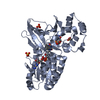
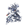
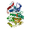
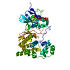
| Title | 1.80 A resolution structure of MAO from P. nicotinovorans in complex with FAD | ||||||
 Components Components | 4-methylaminobutanoate oxidase (methylamine-forming) | ||||||
 Keywords Keywords | OXIDOREDUCTASE / MAO / FAD binding / amine oxidase / flavin oxidase / N-methyl-GABA / GABA | ||||||
| Function / homology | 4-methylaminobutanoate oxidase (methylamine-forming) / nicotine catabolic process / : / Amine oxidase / Flavin containing amine oxidoreductase / FAD/NAD(P)-binding domain superfamily / oxidoreductase activity / FLAVIN-ADENINE DINUCLEOTIDE / 4-methylaminobutanoate oxidase (methylamine-forming) Function and homology information Function and homology information | ||||||
| Biological species |  Paenarthrobacter nicotinovorans (bacteria) Paenarthrobacter nicotinovorans (bacteria) | ||||||
| Method |  X-RAY DIFFRACTION / SYNCHROTRON / SAD / molecular replacement / Resolution: 1.8 Å X-RAY DIFFRACTION / SYNCHROTRON / SAD / molecular replacement / Resolution: 1.8 Å | ||||||
 Authors Authors | Lovell, S. / Bowman, A. / Battaile, K.P. / Deay, D.O. | ||||||
| Funding support | 1items
| ||||||
 Citation Citation | Journal: To be published Title: To be determined Authors: Bowman, A. / Deay, D.O. / Battaile, K.P. / Lovell, S. | ||||||
| History |
|
- Structure visualization
| Structure viewer | Molecule: MolmilJmol/JSmol |
|---|
- Downloads & links
-Download
| PDBx/mmCIF format | 7rt0.cif.gz | 94.7 KB | Display | PDBx/mmCIF format |
|---|---|---|---|---|
| PDB format | pdb7rt0.ent.gz | 68.4 KB | Display | PDB format |
| PDBx/mmJSON format | 7rt0.json.gz | Tree view | PDBx/mmJSON format | |
| Others |  Other downloads Other downloads |
-Validation report
| Arichive directory | https://data.pdbj.org/pub/pdb/validation_reports/rt/7rt0ftp://data.pdbj.org/pub/pdb/validation_reports/rt/7rt0 | HTTPS FTP |
|---|
-Related structure data
| Related structure data | 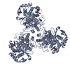 4dyaC 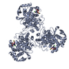 4dybC 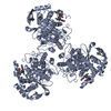 4dynC 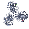 4dypC  4dysC  4dytC  5iboC  6dqcC  6dqdC  6dqeC  6dqfC  9bqnC C: citing same article ( |
|---|---|
| Similar structure data |
-Links
 PDBj
PDBj
- Assembly
| Deposited unit | 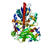
| ||||||||
|---|---|---|---|---|---|---|---|---|---|
| 1 |
| ||||||||
| Unit cell |
| ||||||||
| Components on special symmetry positions |
|
-Components
| #1: Protein | Mass: 44475.883 Da / Num. of mol.: 1 Source method: isolated from a genetically manipulated source Source: (gene. exp.) Paenarthrobacter nicotinovorans (bacteria)Gene: mao, ORF56 / Plasmid: pTBSG / Production host: References: UniProt: Q8GAJ0, 4-methylaminobutanoate oxidase (methylamine-forming) |
|---|---|
| #2: Chemical | ChemComp-FAD /   Mass: 785.550 Da / Num. of mol.: 1 / Source method: obtained synthetically / Formula: C27H33N9O15P2 / Feature type: SUBJECT OF INVESTIGATION / Comment: FAD*YM Mass: 785.550 Da / Num. of mol.: 1 / Source method: obtained synthetically / Formula: C27H33N9O15P2 / Feature type: SUBJECT OF INVESTIGATION / Comment: FAD*YM |
| #3: Chemical | ChemComp-PG4 /   Mass: 194.226 Da / Num. of mol.: 1 / Source method: obtained synthetically / Formula: C8H18O5 / Comment: precipitant*YM Mass: 194.226 Da / Num. of mol.: 1 / Source method: obtained synthetically / Formula: C8H18O5 / Comment: precipitant*YM |
| #4: Water | ChemComp-HOH /  Mass: 18.015 Da / Num. of mol.: 286 / Source method: isolated from a natural source / Formula: H2O Mass: 18.015 Da / Num. of mol.: 286 / Source method: isolated from a natural source / Formula: H2O |
| Has ligand of interest | Y |
-Experimental details
-Experiment
| Experiment | Method: X-RAY DIFFRACTION / Number of used crystals: 1 |
|---|
- Sample preparation
| Crystal | Density Matthews: 2.7 Å3/Da / Density % sol: 54.47 % / Mosaicity: 0.11 ° |
|---|---|
| Crystal grow | Temperature: 298 K / Method: vapor diffusion, sitting drop / pH: 8.2 / Details: 20% (w/v) PEG 6000, 0.1 M Bicine |
-Data collection
| Diffraction | Mean temperature: 100 K / Serial crystal experiment: N | ||||||||||||||||||||||||
|---|---|---|---|---|---|---|---|---|---|---|---|---|---|---|---|---|---|---|---|---|---|---|---|---|---|
| Diffraction source | Source: SYNCHROTRON / Site: APS  / Beamline: 17-ID / Wavelength: 1 Å / Beamline: 17-ID / Wavelength: 1 Å | ||||||||||||||||||||||||
| Detector | Type: DECTRIS PILATUS 6M / Detector: PIXEL / Date: Jun 20, 2021 | ||||||||||||||||||||||||
| Radiation | Protocol: SINGLE WAVELENGTH / Monochromatic (M) / Laue (L): M / Scattering type: x-ray | ||||||||||||||||||||||||
| Radiation wavelength | Wavelength: 1 Å / Relative weight: 1 | ||||||||||||||||||||||||
| Reflection | Resolution: 1.8→45.69 Å / Num. obs: 44295 / % possible obs: 100 % / Redundancy: 6.8 % / Biso Wilson estimate: 18.56 Å2 / CC1/2: 0.995 / Rmerge(I) obs: 0.145 / Net I/σ(I): 8.6 / Num. measured all: 303398 / Scaling rejects: 22 | ||||||||||||||||||||||||
| Reflection shell | Diffraction-ID: 1
|
-Phasing
| Phasing | Method: molecular replacement | |||||||||
|---|---|---|---|---|---|---|---|---|---|---|
| Phasing MR | Model details: Phaser MODE: MR_AUTO
|
- Processing
| Software |
| ||||||||||||||||||||||||||||||||||||||||||||||||||||||||||||||||||||||||||||||||||||||||||||||||||||||
|---|---|---|---|---|---|---|---|---|---|---|---|---|---|---|---|---|---|---|---|---|---|---|---|---|---|---|---|---|---|---|---|---|---|---|---|---|---|---|---|---|---|---|---|---|---|---|---|---|---|---|---|---|---|---|---|---|---|---|---|---|---|---|---|---|---|---|---|---|---|---|---|---|---|---|---|---|---|---|---|---|---|---|---|---|---|---|---|---|---|---|---|---|---|---|---|---|---|---|---|---|---|---|---|
| Refinement | Method to determine structure: SAD / Resolution: 1.8→45.69 Å / SU ML: 0.17 / Cross valid method: THROUGHOUT / σ(F): 1.02 / Phase error: 18.1 / Stereochemistry target values: ML
| ||||||||||||||||||||||||||||||||||||||||||||||||||||||||||||||||||||||||||||||||||||||||||||||||||||||
| Solvent computation | Shrinkage radii: 0.9 Å / VDW probe radii: 1.11 Å / Solvent model: FLAT BULK SOLVENT MODEL | ||||||||||||||||||||||||||||||||||||||||||||||||||||||||||||||||||||||||||||||||||||||||||||||||||||||
| Displacement parameters | Biso max: 96.48 Å2 / Biso mean: 23.1997 Å2 / Biso min: 8.95 Å2 | ||||||||||||||||||||||||||||||||||||||||||||||||||||||||||||||||||||||||||||||||||||||||||||||||||||||
| Refinement step | Cycle: final / Resolution: 1.8→45.69 Å
| ||||||||||||||||||||||||||||||||||||||||||||||||||||||||||||||||||||||||||||||||||||||||||||||||||||||
| LS refinement shell | Refine-ID: X-RAY DIFFRACTION / Rfactor Rfree error: 0 / Total num. of bins used: 16 / % reflection obs: 100 %
|
