 Movie
Movie Controller

[English] 日本語
 Yorodumi
- PDB-6x44: High Resolution Crystal Structure Analysis of SERA5 proenzyme fro... -
+ Open data

- Basic information
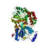
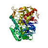
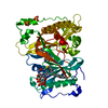
| Entry | Database: PDB / ID: 6x44 | ||||||
|---|---|---|---|---|---|---|---|
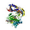
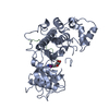
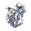
| Title | High Resolution Crystal Structure Analysis of SERA5 proenzyme from plasmodium falciparum | ||||||
 Components Components | Serine repeat antigen 5 | ||||||
 Keywords Keywords | HYDROLASE / malaria / prodomain / protease | ||||||
| Function / homology |  Function and homology information Function and homology informationmembrane-bounded organelle / symbiont-containing vacuolar space / exit from host cell / symbiont-containing vacuole / regulation of immune response / cysteine-type peptidase activity / serine-type peptidase activity / kinase binding / peptidase activity / lysosome ...membrane-bounded organelle / symbiont-containing vacuolar space / exit from host cell / symbiont-containing vacuole / regulation of immune response / cysteine-type peptidase activity / serine-type peptidase activity / kinase binding / peptidase activity / lysosome / Hydrolases; Acting on peptide bonds (peptidases); Cysteine endopeptidases / cysteine-type endopeptidase activity / proteolysis / : / plasma membrane Similarity search - Function | ||||||
| Biological species |  | ||||||
| Method |  X-RAY DIFFRACTION / SYNCHROTRON / MOLECULAR REPLACEMENT / Resolution: 2.19733954166 Å X-RAY DIFFRACTION / SYNCHROTRON / MOLECULAR REPLACEMENT / Resolution: 2.19733954166 Å | ||||||
 Authors Authors | Clarke, O.B. / Smith, N.A. / Lee, M. / Smith, B.J. | ||||||
 Citation Citation | Journal: Protein Sci. / Year: 2020 Title: Structure of the Plasmodium falciparum PfSERA5 pseudo-zymogen. Authors: Smith, N.A. / Clarke, O.B. / Lee, M. / Hodder, A.N. / Smith, B.J. | ||||||
| History |
|
- Structure visualization
| Structure viewer | Molecule: MolmilJmol/JSmol |
|---|
- Downloads & links
-Download
| PDBx/mmCIF format | 6x44.cif.gz | 361.7 KB | Display | PDBx/mmCIF format |
|---|---|---|---|---|
| PDB format | pdb6x44.ent.gz | 238.4 KB | Display | PDB format |
| PDBx/mmJSON format | 6x44.json.gz | Tree view | PDBx/mmJSON format | |
| Others |  Other downloads Other downloads |
-Validation report
| Arichive directory | https://data.pdbj.org/pub/pdb/validation_reports/x4/6x44ftp://data.pdbj.org/pub/pdb/validation_reports/x4/6x44 | HTTPS FTP |
|---|
-Related structure data
| Related structure data | 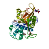 6x42C 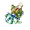 2wbfS S: Starting model for refinement C: citing same article ( |
|---|---|
| Similar structure data |


-Links
 PDBj
PDBj
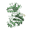
- Assembly
| Deposited unit | 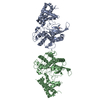
| ||||||||||||
|---|---|---|---|---|---|---|---|---|---|---|---|---|---|
| 1 | 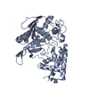
| ||||||||||||
| 2 | 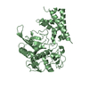
| ||||||||||||
| Unit cell |
|
-Components
| #1: Protein | Mass: 50848.332 Da / Num. of mol.: 2 Source method: isolated from a genetically manipulated source Source: (gene. exp.) Gene: sera5 / Production host:  #2: Chemical | ChemComp-PEG / |   Mass: 106.120 Da / Num. of mol.: 1 / Source method: obtained synthetically / Formula: C4H10O3 Mass: 106.120 Da / Num. of mol.: 1 / Source method: obtained synthetically / Formula: C4H10O3#3: Water | ChemComp-HOH / |  Mass: 18.015 Da / Num. of mol.: 230 / Source method: isolated from a natural source / Formula: H2O Mass: 18.015 Da / Num. of mol.: 230 / Source method: isolated from a natural source / Formula: H2OHas ligand of interest | N | Has protein modification | Y | |
|---|
-Experimental details
-Experiment
| Experiment | Method: X-RAY DIFFRACTION / Number of used crystals: 1 |
|---|
- Sample preparation
| Crystal | Density Matthews: 2.69 Å3/Da / Density % sol: 54.33 % |
|---|---|
| Crystal grow | Temperature: 293 K / Method: vapor diffusion, hanging drop / pH: 8.1 / Details: LiSO4, Tris-HCl, PEG-4000 |
-Data collection
| Diffraction | Mean temperature: 100 K / Serial crystal experiment: N |
|---|---|
| Diffraction source | Source: SYNCHROTRON / Site: APS  / Beamline: 24-ID-C / Wavelength: 1.0393 Å / Beamline: 24-ID-C / Wavelength: 1.0393 Å |
| Detector | Type: DECTRIS PILATUS 6M-F / Detector: PIXEL / Date: Jul 18, 2013 |
| Radiation | Protocol: SINGLE WAVELENGTH / Monochromatic (M) / Laue (L): M / Scattering type: x-ray |
| Radiation wavelength | Wavelength: 1.0393 Å / Relative weight: 1 |
| Reflection | Resolution: 2.197→47.55 Å / Num. obs: 56459 / % possible obs: 99.07 % / Redundancy: 7.3 % / Biso Wilson estimate: 34.5665711195 Å2 / CC1/2: 0.998 / Rmerge(I) obs: 0.1162 / Rpim(I) all: 0.04576 / Rrim(I) all: 0.1252 / Net I/σ(I): 11.56 |
| Reflection shell | Resolution: 2.197→2.276 Å / Redundancy: 7.5 % / Rmerge(I) obs: 0.9169 / Num. unique obs: 5430 / CC1/2: 0.737 / Rpim(I) all: 0.3555 / Rrim(I) all: 0.9851 / % possible all: 97.12 |
- Processing
| Software |
| |||||||||||||||||||||||||||||||||||||||||||||||||||||||||||||||||||||||||||||||||||||||||||||||||||||||||||||||||||||||||||||||||||||||||||||||||||
|---|---|---|---|---|---|---|---|---|---|---|---|---|---|---|---|---|---|---|---|---|---|---|---|---|---|---|---|---|---|---|---|---|---|---|---|---|---|---|---|---|---|---|---|---|---|---|---|---|---|---|---|---|---|---|---|---|---|---|---|---|---|---|---|---|---|---|---|---|---|---|---|---|---|---|---|---|---|---|---|---|---|---|---|---|---|---|---|---|---|---|---|---|---|---|---|---|---|---|---|---|---|---|---|---|---|---|---|---|---|---|---|---|---|---|---|---|---|---|---|---|---|---|---|---|---|---|---|---|---|---|---|---|---|---|---|---|---|---|---|---|---|---|---|---|---|---|---|---|
| Refinement | Method to determine structure: MOLECULAR REPLACEMENT Starting model: 2wbf Resolution: 2.19733954166→47.5499422318 Å / SU ML: 0.238223530532 / Cross valid method: FREE R-VALUE / σ(F): 1.34004080654 / Phase error: 21.762703983 Stereochemistry target values: GeoStd + Monomer Library + CDL v1.2
| |||||||||||||||||||||||||||||||||||||||||||||||||||||||||||||||||||||||||||||||||||||||||||||||||||||||||||||||||||||||||||||||||||||||||||||||||||
| Solvent computation | Shrinkage radii: 0.9 Å / VDW probe radii: 1.11 Å / Solvent model: FLAT BULK SOLVENT MODEL | |||||||||||||||||||||||||||||||||||||||||||||||||||||||||||||||||||||||||||||||||||||||||||||||||||||||||||||||||||||||||||||||||||||||||||||||||||
| Displacement parameters | Biso mean: 46.2048237869 Å2 | |||||||||||||||||||||||||||||||||||||||||||||||||||||||||||||||||||||||||||||||||||||||||||||||||||||||||||||||||||||||||||||||||||||||||||||||||||
| Refinement step | Cycle: LAST / Resolution: 2.19733954166→47.5499422318 Å
| |||||||||||||||||||||||||||||||||||||||||||||||||||||||||||||||||||||||||||||||||||||||||||||||||||||||||||||||||||||||||||||||||||||||||||||||||||
| Refine LS restraints |
| |||||||||||||||||||||||||||||||||||||||||||||||||||||||||||||||||||||||||||||||||||||||||||||||||||||||||||||||||||||||||||||||||||||||||||||||||||
| LS refinement shell |
|
