 Movie
Movie Controller

[English] 日本語
 Yorodumi
- PDB-2wbf: Crystal Structure Analysis of SERA5E from plasmodium falciparum w... -
+ Open data

- Basic information
| Entry | Database: PDB / ID: 2wbf | |||||||||
|---|---|---|---|---|---|---|---|---|---|---|
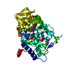
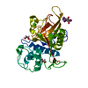
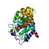
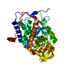
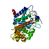
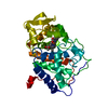
| Title | Crystal Structure Analysis of SERA5E from plasmodium falciparum with loop 690-700 ordered | |||||||||
 Components Components | SERINE-REPEAT ANTIGEN PROTEIN | |||||||||
 Keywords Keywords | HYDROLASE / SERINE REPEAT ANTIGEN / SERA / MALARIA / VACUOLE / PROTEASE / CATHEPSIN / PLASMODIUM / GLYCOPROTEIN / THIOL PROTEASE | |||||||||
| Function / homology |  Function and homology information Function and homology informationsymbiont-containing vacuolar space / cysteine-type peptidase activity / Hydrolases; Acting on peptide bonds (peptidases); Cysteine endopeptidases / proteolysis / extracellular region / plasma membrane Similarity search - Function | |||||||||
| Biological species |  | |||||||||
| Method |  X-RAY DIFFRACTION / SYNCHROTRON / MOLECULAR REPLACEMENT / Resolution: 1.6 Å X-RAY DIFFRACTION / SYNCHROTRON / MOLECULAR REPLACEMENT / Resolution: 1.6 Å | |||||||||
 Authors Authors | Smith, B.J. / Malby, R.L. / Colman, P.M. / Clarke, O.B. | |||||||||
 Citation Citation | Journal: J. Mol. Biol. / Year: 2009 Title: Structural insights into the protease-like antigen Plasmodium falciparum SERA5 and its noncanonical active-site serine. Authors: Hodder, A.N. / Malby, R.L. / Clarke, O.B. / Fairlie, W.D. / Colman, P.M. / Crabb, B.S. / Smith, B.J. | |||||||||
| History |
|
- Structure visualization
| Structure viewer | Molecule: MolmilJmol/JSmol |
|---|
- Downloads & links
-Download
| PDBx/mmCIF format | 2wbf.cif.gz | 169.4 KB | Display | PDBx/mmCIF format |
|---|---|---|---|---|
| PDB format | pdb2wbf.ent.gz | 135.5 KB | Display | PDB format |
| PDBx/mmJSON format | 2wbf.json.gz | Tree view | PDBx/mmJSON format | |
| Others |  Other downloads Other downloads |
-Validation report
| Arichive directory | https://data.pdbj.org/pub/pdb/validation_reports/wb/2wbfftp://data.pdbj.org/pub/pdb/validation_reports/wb/2wbf | HTTPS FTP |
|---|
-Related structure data
| Related structure data | 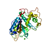 3ch2SC 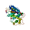 3ch3C S: Starting model for refinement C: citing same article ( |
|---|---|
| Similar structure data |

-Links
 PDBj
PDBj
- Assembly
| Deposited unit | 
| ||||||||
|---|---|---|---|---|---|---|---|---|---|
| 1 |
| ||||||||
| Unit cell |
|
-Components
| #1: Protein | Mass: 30666.279 Da / Num. of mol.: 1 / Fragment: SERINE-PROTEASE DOMAIN, RESIDUES 555-819 Source method: isolated from a genetically manipulated source Source: (gene. exp.) Production host:  | ||||||||
|---|---|---|---|---|---|---|---|---|---|
| #2: Chemical | ChemComp-DMS /   Mass: 78.133 Da / Num. of mol.: 6 / Source method: obtained synthetically / Formula: C2H6OS / Comment: DMSO, precipitant*YM Mass: 78.133 Da / Num. of mol.: 6 / Source method: obtained synthetically / Formula: C2H6OS / Comment: DMSO, precipitant*YM#3: Chemical |   Mass: 40.078 Da / Num. of mol.: 2 / Source method: obtained synthetically / Formula: Ca Mass: 40.078 Da / Num. of mol.: 2 / Source method: obtained synthetically / Formula: Ca#4: Chemical | ChemComp-CL /   Mass: 35.453 Da / Num. of mol.: 4 / Source method: obtained synthetically / Formula: Cl Mass: 35.453 Da / Num. of mol.: 4 / Source method: obtained synthetically / Formula: Cl#5: Water | ChemComp-HOH / |  Mass: 18.015 Da / Num. of mol.: 248 / Source method: isolated from a natural source / Formula: H2O Mass: 18.015 Da / Num. of mol.: 248 / Source method: isolated from a natural source / Formula: H2OHas protein modification | Y | |
-Experimental details
-Experiment
| Experiment | Method: X-RAY DIFFRACTION / Number of used crystals: 1 |
|---|
- Sample preparation
| Crystal | Density Matthews: 2.5 Å3/Da / Density % sol: 50 % / Description: NONE |
|---|---|
| Crystal grow | Method: vapor diffusion, sitting drop / pH: 6.5 Details: 17% PEG3350,0.2M CACL2, 5% DMSO, 10MM BIS-TRIS PH 6.5, 10MM NACL, SITTING-DROP VAPOUR-DIFFUSION |
-Data collection
| Diffraction | Mean temperature: 100 K |
|---|---|
| Diffraction source | Source: SYNCHROTRON / Site: SLS  / Beamline: X06SA / Wavelength: 0.95667 / Beamline: X06SA / Wavelength: 0.95667 |
| Detector | Type: MARRESEARCH / Detector: CCD / Date: Jun 21, 2008 |
| Radiation | Protocol: SINGLE WAVELENGTH / Monochromatic (M) / Laue (L): M / Scattering type: x-ray |
| Radiation wavelength | Wavelength: 0.95667 Å / Relative weight: 1 |
| Reflection | Resolution: 1.6→30 Å / Num. obs: 36811 / % possible obs: 99.7 % / Observed criterion σ(I): 0 / Redundancy: 5.5 % / Biso Wilson estimate: 16.63 Å2 / Rmerge(I) obs: 0.07 / Net I/σ(I): 24.6 |
| Reflection shell | Resolution: 1.6→1.66 Å / Redundancy: 5.1 % / Rmerge(I) obs: 0.67 / Mean I/σ(I) obs: 2.6 / % possible all: 100 |
- Processing
| Software |
| ||||||||||||||||||||||||||||||||||||||||||||||||||||||||||||||||||||||||||||||||||||||||||||||||||
|---|---|---|---|---|---|---|---|---|---|---|---|---|---|---|---|---|---|---|---|---|---|---|---|---|---|---|---|---|---|---|---|---|---|---|---|---|---|---|---|---|---|---|---|---|---|---|---|---|---|---|---|---|---|---|---|---|---|---|---|---|---|---|---|---|---|---|---|---|---|---|---|---|---|---|---|---|---|---|---|---|---|---|---|---|---|---|---|---|---|---|---|---|---|---|---|---|---|---|---|
| Refinement | Method to determine structure: MOLECULAR REPLACEMENT Starting model: PDB ENTRY 3CH2 Resolution: 1.6→29.55 Å / SU ML: 0.67 / σ(F): 1.96 / Phase error: 16.78 / Stereochemistry target values: ML
| ||||||||||||||||||||||||||||||||||||||||||||||||||||||||||||||||||||||||||||||||||||||||||||||||||
| Solvent computation | Shrinkage radii: 0.9 Å / VDW probe radii: 1.11 Å / Solvent model: FLAT BULK SOLVENT MODEL / Bsol: 56.89 Å2 / ksol: 0.43 e/Å3 | ||||||||||||||||||||||||||||||||||||||||||||||||||||||||||||||||||||||||||||||||||||||||||||||||||
| Displacement parameters |
| ||||||||||||||||||||||||||||||||||||||||||||||||||||||||||||||||||||||||||||||||||||||||||||||||||
| Refinement step | Cycle: LAST / Resolution: 1.6→29.55 Å
| ||||||||||||||||||||||||||||||||||||||||||||||||||||||||||||||||||||||||||||||||||||||||||||||||||
| Refine LS restraints |
| ||||||||||||||||||||||||||||||||||||||||||||||||||||||||||||||||||||||||||||||||||||||||||||||||||
| LS refinement shell |
| ||||||||||||||||||||||||||||||||||||||||||||||||||||||||||||||||||||||||||||||||||||||||||||||||||
| Refinement TLS params. | Method: refined / Refine-ID: X-RAY DIFFRACTION
| ||||||||||||||||||||||||||||||||||||||||||||||||||||||||||||||||||||||||||||||||||||||||||||||||||
| Refinement TLS group |
|
