| Entry | Database: PDB / ID: 5f84
|
|---|
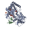
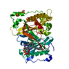
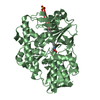
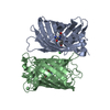
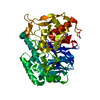
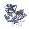
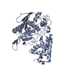
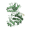
| Title | Crystal structure of Drosophila Poglut1 (Rumi) complexed with its glycoprotein product (glucosylated EGF repeat) and UDP |
|---|
 Components Components | - Coagulation factor IX
- O-glucosyltransferase rumi
|
|---|
 Keywords Keywords | Transferase/Hydrolase / glycosyltransferase / protein O-glucosyltransferase / Notch regulation / EGF repeat / Transferase-Hydrolase complex |
|---|
| Function / homology |  Function and homology information Function and homology information
EGF-domain serine glucosyltransferase activity / EGF-domain serine xylosyltransferase activity / muscle tissue development / protein O-linked glycosylation via glucose / rhabdomere development / UDP-xylosyltransferase activity / Defective F9 secretion / coagulation factor IXa / UDP-glucosyltransferase activity / Defective gamma-carboxylation of F9 ...EGF-domain serine glucosyltransferase activity / EGF-domain serine xylosyltransferase activity / muscle tissue development / protein O-linked glycosylation via glucose / rhabdomere development / UDP-xylosyltransferase activity / Defective F9 secretion / coagulation factor IXa / UDP-glucosyltransferase activity / Defective gamma-carboxylation of F9 / glucosyltransferase activity / Defective F9 activation / protein O-linked glycosylation / Defective factor IX causes thrombophilia / Defective cofactor function of FVIIIa variant / Defective F9 variant does not activate FX / : / zymogen activation / Transferases; Glycosyltransferases; Hexosyltransferases / positive regulation of Notch signaling pathway / negative regulation of Notch signaling pathway / Protein hydroxylation / cell fate commitment / Transport of gamma-carboxylated protein precursors from the endoplasmic reticulum to the Golgi apparatus / Gamma-carboxylation of protein precursors / Removal of aminoterminal propeptides from gamma-carboxylated proteins / Notch signaling pathway / : / endomembrane system / Golgi lumen / blood coagulation / extracellular matrix / endopeptidase activity / endoplasmic reticulum lumen / serine-type endopeptidase activity / calcium ion binding / proteolysis / : / extracellular exosome / extracellular region / metal ion binding / plasma membraneSimilarity search - Function Glycosyl transferase CAP10 domain / : / Glycosyl transferase family 90 / Putative lipopolysaccharide-modifying enzyme. / Endoplasmic reticulum targeting sequence. / Peptidase S1A, coagulation factor VII/IX/X/C/Z / : / Coagulation factor-like, Gla domain superfamily / Coagulation Factor Xa inhibitory site / EGF-like domain ...Glycosyl transferase CAP10 domain / : / Glycosyl transferase family 90 / Putative lipopolysaccharide-modifying enzyme. / Endoplasmic reticulum targeting sequence. / Peptidase S1A, coagulation factor VII/IX/X/C/Z / : / Coagulation factor-like, Gla domain superfamily / Coagulation Factor Xa inhibitory site / EGF-like domain / EGF-type aspartate/asparagine hydroxylation site / EGF-like calcium-binding, conserved site / Calcium-binding EGF-like domain signature. / Aspartic acid and asparagine hydroxylation site. / EGF-like calcium-binding domain / Calcium-binding EGF-like domain / Vitamin K-dependent carboxylation/gamma-carboxyglutamic (GLA) domain / Gamma-carboxyglutamic acid-rich (GLA) domain / Gamma-carboxyglutamic acid-rich (GLA) domain superfamily / Vitamin K-dependent carboxylation domain. / Gla domain profile. / Domain containing Gla (gamma-carboxyglutamate) residues. / Epidermal growth factor-like domain. / EGF-like domain profile. / EGF-like domain signature 1. / EGF-like domain signature 2. / EGF-like domain / Serine proteases, trypsin family, histidine active site / Serine proteases, trypsin family, serine active site / Serine proteases, trypsin family, histidine active site. / Serine proteases, trypsin family, serine active site. / Peptidase S1A, chymotrypsin family / Serine proteases, trypsin domain profile. / Trypsin-like serine protease / Serine proteases, trypsin domain / Trypsin / Peptidase S1, PA clan, chymotrypsin-like fold / Peptidase S1, PA clanSimilarity search - Domain/homology |
|---|
| Biological species |   Drosophila melanogaster (fruit fly) Drosophila melanogaster (fruit fly)
 Homo sapiens (human) Homo sapiens (human) |
|---|
| Method |  X-RAY DIFFRACTION / SYNCHROTRON / MOLECULAR REPLACEMENT / Resolution: 2.5 Å X-RAY DIFFRACTION / SYNCHROTRON / MOLECULAR REPLACEMENT / Resolution: 2.5 Å |
|---|
 Authors Authors | Yu, H.J. / Li, H.L. |
|---|
 Citation Citation | Journal: Nat. Chem. Biol. / Year: 2016
Title: Structural analysis of Notch-regulating Rumi reveals basis for pathogenic mutations.
Authors: Yu, H. / Takeuchi, H. / Takeuchi, M. / Liu, Q. / Kantharia, J. / Haltiwanger, R.S. / Li, H. |
|---|
| History | | Deposition | Dec 9, 2015 | Deposition site: RCSB / Processing site: RCSB |
|---|
| Revision 1.0 | Jul 20, 2016 | Provider: repository / Type: Initial release |
|---|
| Revision 1.1 | Apr 18, 2018 | Group: Data collection / Database references / Derived calculations
Category: citation / citation_author / pdbx_struct_oper_list
Item: _citation.country / _citation.journal_abbrev ..._citation.country / _citation.journal_abbrev / _citation.journal_id_CSD / _citation.journal_id_ISSN / _citation.journal_volume / _citation.page_first / _citation.page_last / _citation.pdbx_database_id_DOI / _citation.pdbx_database_id_PubMed / _citation.title / _citation.year / _pdbx_struct_oper_list.symmetry_operation |
|---|
| Revision 1.2 | Jul 29, 2020 | Group: Data collection / Derived calculations / Structure summary
Category: chem_comp / entity ...chem_comp / entity / pdbx_chem_comp_identifier / pdbx_entity_nonpoly / struct_conn / struct_site / struct_site_gen
Item: _chem_comp.name / _chem_comp.type ..._chem_comp.name / _chem_comp.type / _entity.pdbx_description / _pdbx_entity_nonpoly.name / _struct_conn.pdbx_role
Description: Carbohydrate remediation / Provider: repository / Type: Remediation |
|---|
| Revision 1.3 | Oct 23, 2024 | Group: Data collection / Database references / Structure summary
Category: chem_comp / chem_comp_atom ...chem_comp / chem_comp_atom / chem_comp_bond / database_2 / pdbx_entry_details / pdbx_modification_feature
Item: _chem_comp.pdbx_synonyms / _database_2.pdbx_DOI / _database_2.pdbx_database_accession |
|---|
|
|---|
 Movie
Movie Controller

 Yorodumi
 Open data
 Basic information
 Structure visualization
 Downloads & links
 Other downloads


 PDBj
PDBj


 Assembly
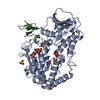


 Type: D-saccharide, beta linking / Mass: 180.156 Da / Num. of mol.: 1


 Type: RNA linking / Mass: 404.161 Da / Num. of mol.: 1 / Source method: obtained synthetically / Formula: C9H14N2O12P2 / Comment: UDP*YM
 Mass: 96.063 Da / Num. of mol.: 5 / Source method: obtained synthetically / Formula: SO4
 Sample preparation
 / Beamline: X25 / Wavelength: 1.1 Å
 Processing