 Movie
Movie Controller

[English] 日本語
 Yorodumi
- PDB-5zca: Crystal structure of lambda repressor (1-20) fused with maltose-b... -
+ Open data

- Basic information
| Entry | Database: PDB / ID: 5zca | |||||||||
|---|---|---|---|---|---|---|---|---|---|---|
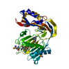
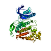
| Title | Crystal structure of lambda repressor (1-20) fused with maltose-binding protein | |||||||||
 Components Components | Repressor protein cI,Maltose-binding periplasmic protein | |||||||||
 Keywords Keywords | DNA BINDING PROTEIN / LAMBDA REPRESSOR / MALTOSE-BINDING PROTEIN / SUGAR BINDING PROTEIN | |||||||||
| Function / homology |  Function and homology information Function and homology informationmaintenance of viral latency / latency-replication decision / positive regulation of viral transcription / : / detection of maltose stimulus / maltose transport complex / carbohydrate transport / carbohydrate transmembrane transporter activity / maltose binding / maltose transport ...maintenance of viral latency / latency-replication decision / positive regulation of viral transcription / : / detection of maltose stimulus / maltose transport complex / carbohydrate transport / carbohydrate transmembrane transporter activity / maltose binding / maltose transport / maltodextrin transmembrane transport / core promoter sequence-specific DNA binding / ATP-binding cassette (ABC) transporter complex, substrate-binding subunit-containing / ATP-binding cassette (ABC) transporter complex / cell chemotaxis / outer membrane-bounded periplasmic space / periplasmic space / DNA damage response / membrane / identical protein binding Similarity search - Function | |||||||||
| Biological species |  Escherichia phage lambda (virus) Escherichia phage lambda (virus) | |||||||||
| Method |  X-RAY DIFFRACTION / SYNCHROTRON / MOLECULAR REPLACEMENT / Resolution: 1.801 Å X-RAY DIFFRACTION / SYNCHROTRON / MOLECULAR REPLACEMENT / Resolution: 1.801 Å | |||||||||
 Authors Authors | Hanazono, Y. / Takeda, K. / Miki, K. | |||||||||
 Citation Citation | Journal: Febs Open Bio / Year: 2018 Title: Co-translational folding of alpha-helical proteins: structural studies of intermediate-length variants of the lambda repressor Authors: Hanazono, Y. / Takeda, K. / Miki, K. | |||||||||
| History |
|
- Structure visualization
| Structure viewer | Molecule: MolmilJmol/JSmol |
|---|
- Downloads & links
-Download
| PDBx/mmCIF format | 5zca.cif.gz | 101.6 KB | Display | PDBx/mmCIF format |
|---|---|---|---|---|
| PDB format | pdb5zca.ent.gz | 74.2 KB | Display | PDB format |
| PDBx/mmJSON format | 5zca.json.gz | Tree view | PDBx/mmJSON format | |
| Others |  Other downloads Other downloads |
-Validation report
| Arichive directory | https://data.pdbj.org/pub/pdb/validation_reports/zc/5zcaftp://data.pdbj.org/pub/pdb/validation_reports/zc/5zca | HTTPS FTP |
|---|
-Related structure data
| Related structure data |  1anfS S: Starting model for refinement |
|---|---|
| Similar structure data |


-Links
 PDBj
PDBj


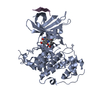
- Assembly
| Deposited unit | 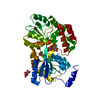
| ||||||||
|---|---|---|---|---|---|---|---|---|---|
| 1 |
| ||||||||
| Unit cell |
|
-Components
| #1: Protein | Mass: 43027.699 Da / Num. of mol.: 1 Source method: isolated from a genetically manipulated source Details: The fusion protein of Repressor protein (residues 1-20), linker, and Maltose-binding periplasmic protein (residues 24-389) Source: (gene. exp.) Escherichia phage lambda (virus), (gene. exp.) Gene: cI, lambdap88, malE, b4034, JW3994 / Strain: K12 / Production host: |
|---|---|
| #2: Polysaccharide | alpha-D-glucopyranose-(1-4)-alpha-D-glucopyranose / alpha-maltose  Source method: isolated from a genetically manipulated source Details: oligosaccharide / References: alpha-maltose |
| #3: Chemical | ChemComp-CIT /   Mass: 192.124 Da / Num. of mol.: 1 / Source method: obtained synthetically / Formula: C6H8O7 Mass: 192.124 Da / Num. of mol.: 1 / Source method: obtained synthetically / Formula: C6H8O7 |
| #4: Water | ChemComp-HOH /  Mass: 18.015 Da / Num. of mol.: 360 / Source method: isolated from a natural source / Formula: H2O Mass: 18.015 Da / Num. of mol.: 360 / Source method: isolated from a natural source / Formula: H2O |
-Experimental details
-Experiment
| Experiment | Method: X-RAY DIFFRACTION / Number of used crystals: 1 |
|---|
- Sample preparation
| Crystal | Density Matthews: 2.07 Å3/Da / Density % sol: 40.52 % |
|---|---|
| Crystal grow | Temperature: 293 K / Method: vapor diffusion, sitting drop / Details: 1.6 M triammonium citrate |
-Data collection
| Diffraction | Mean temperature: 100 K |
|---|---|
| Diffraction source | Source: SYNCHROTRON / Site: SPring-8  / Beamline: BL41XU / Wavelength: 1 Å / Beamline: BL41XU / Wavelength: 1 Å |
| Detector | Type: DECTRIS PILATUS3 6M / Detector: PIXEL / Date: Apr 19, 2017 |
| Radiation | Protocol: SINGLE WAVELENGTH / Monochromatic (M) / Laue (L): M / Scattering type: x-ray |
| Radiation wavelength | Wavelength: 1 Å / Relative weight: 1 |
| Reflection | Resolution: 1.8→50 Å / Num. obs: 33855 / % possible obs: 100 % / Redundancy: 6.4 % / Rsym value: 0.083 / Net I/σ(I): 17.5 |
| Reflection shell | Resolution: 1.8→1.83 Å / Redundancy: 6.2 % / Mean I/σ(I) obs: 1.6 / Rsym value: 0.817 / % possible all: 100 |
- Processing
| Software |
| |||||||||||||||||||||||||||||||||||||||||||||||||||||||||||||||||||||||||||||||||||||||||||
|---|---|---|---|---|---|---|---|---|---|---|---|---|---|---|---|---|---|---|---|---|---|---|---|---|---|---|---|---|---|---|---|---|---|---|---|---|---|---|---|---|---|---|---|---|---|---|---|---|---|---|---|---|---|---|---|---|---|---|---|---|---|---|---|---|---|---|---|---|---|---|---|---|---|---|---|---|---|---|---|---|---|---|---|---|---|---|---|---|---|---|---|---|
| Refinement | Method to determine structure: MOLECULAR REPLACEMENT Starting model: 1ANF Resolution: 1.801→45.566 Å / SU ML: 0.21 / Cross valid method: FREE R-VALUE / σ(F): 1.37 / Phase error: 20.13
| |||||||||||||||||||||||||||||||||||||||||||||||||||||||||||||||||||||||||||||||||||||||||||
| Solvent computation | Shrinkage radii: 0.9 Å / VDW probe radii: 1.11 Å | |||||||||||||||||||||||||||||||||||||||||||||||||||||||||||||||||||||||||||||||||||||||||||
| Refinement step | Cycle: LAST / Resolution: 1.801→45.566 Å
| |||||||||||||||||||||||||||||||||||||||||||||||||||||||||||||||||||||||||||||||||||||||||||
| Refine LS restraints |
| |||||||||||||||||||||||||||||||||||||||||||||||||||||||||||||||||||||||||||||||||||||||||||
| LS refinement shell |
|
