negative regulation of glycogen (starch) synthase activity / neuron projection organization / regulation of microtubule anchoring at centrosome / negative regulation of mesenchymal stem cell differentiation / negative regulation of type B pancreatic cell development / superior temporal gyrus development / positive regulation of protein localization to cilium / negative regulation of glycogen biosynthetic process / negative regulation of TORC2 signaling / beta-arrestin-dependent dopamine receptor signaling pathway ...negative regulation of glycogen (starch) synthase activity / neuron projection organization / regulation of microtubule anchoring at centrosome / negative regulation of mesenchymal stem cell differentiation / negative regulation of type B pancreatic cell development / superior temporal gyrus development / positive regulation of protein localization to cilium / negative regulation of glycogen biosynthetic process / negative regulation of TORC2 signaling / beta-arrestin-dependent dopamine receptor signaling pathway / negative regulation of dopaminergic neuron differentiation / positive regulation of protein localization to centrosome / maintenance of cell polarity / regulation of protein export from nucleus / positive regulation of cilium assembly / heart valve development / CRMPs in Sema3A signaling / tau-protein kinase / beta-catenin destruction complex / APC truncation mutants have impaired AXIN binding / AXIN missense mutants destabilize the destruction complex / Truncations of AMER1 destabilize the destruction complex / positive regulation of mitochondrial outer membrane permeabilization involved in apoptotic signaling pathway / cellular response to interleukin-3 / Maturation of nucleoprotein / Beta-catenin phosphorylation cascade / Signaling by GSK3beta mutants / CTNNB1 S33 mutants aren't phosphorylated / CTNNB1 S37 mutants aren't phosphorylated / CTNNB1 S45 mutants aren't phosphorylated / CTNNB1 T41 mutants aren't phosphorylated / negative regulation of TOR signaling / regulation of long-term synaptic potentiation / Wnt signalosome / regulation of microtubule-based process / AKT phosphorylates targets in the cytosol / Disassembly of the destruction complex and recruitment of AXIN to the membrane / positive regulation of protein binding / regulation of axon extension / negative regulation of calcineurin-NFAT signaling cascade / negative regulation of protein localization to nucleus / Maturation of nucleoprotein / negative regulation of epithelial to mesenchymal transition / tau-protein kinase activity / positive regulation of cell-matrix adhesion / glycogen metabolic process / ER overload response / regulation of axonogenesis / regulation of dendrite morphogenesis / regulation of neuron projection development / establishment of cell polarity / Constitutive Signaling by AKT1 E17K in Cancer / protein kinase A catalytic subunit binding / dynactin binding / epithelial to mesenchymal transition / canonical Wnt signaling pathway / Regulation of HSF1-mediated heat shock response / negative regulation of osteoblast differentiation / NF-kappaB binding / negative regulation of extrinsic apoptotic signaling pathway via death domain receptors / extrinsic apoptotic signaling pathway / negative regulation of protein-containing complex assembly / regulation of cellular response to heat / cellular response to retinoic acid / extrinsic apoptotic signaling pathway in absence of ligand / positive regulation of type I interferon production / Transcriptional and post-translational regulation of MITF-M expression and activity / positive regulation of autophagy / response to endoplasmic reticulum stress / positive regulation of protein export from nucleus / presynaptic modulation of chemical synaptic transmission / negative regulation of cell migration / excitatory postsynaptic potential / positive regulation of protein ubiquitination / peptidyl-serine phosphorylation / hippocampus development / regulation of microtubule cytoskeleton organization / positive regulation of cell differentiation / mitochondrion organization / Ubiquitin-dependent degradation of Cyclin D / negative regulation of canonical Wnt signaling pathway / circadian rhythm / positive regulation of protein-containing complex assembly / regulation of circadian rhythm / GSK3B and BTRC:CUL1-mediated-degradation of NFE2L2 / beta-catenin binding / B-WICH complex positively regulates rRNA expression / Degradation of GLI2 by the proteasome / GLI3 is processed to GLI3R by the proteasome / tau protein binding / Degradation of beta-catenin by the destruction complex / cellular response to amyloid-beta / Wnt signaling pathway / neuron projection development / kinase activity / positive regulation of protein catabolic process / p53 binding / Regulation of RUNX2 expression and activity / insulin receptor signaling pathway / protein autophosphorylation Similarity search - Function
Glycogen synthase kinase 3, catalytic domain / : / Phosphorylase Kinase; domain 1 / Phosphorylase Kinase; domain 1 / Transferase(Phosphotransferase) domain 1 / Transferase(Phosphotransferase); domain 1 / Serine/threonine-protein kinase, active site / Serine/Threonine protein kinases active-site signature. / Protein kinase domain / Serine/Threonine protein kinases, catalytic domain ...Glycogen synthase kinase 3, catalytic domain / : / Phosphorylase Kinase; domain 1 / Phosphorylase Kinase; domain 1 / Transferase(Phosphotransferase) domain 1 / Transferase(Phosphotransferase); domain 1 / Serine/threonine-protein kinase, active site / Serine/Threonine protein kinases active-site signature. / Protein kinase domain / Serine/Threonine protein kinases, catalytic domain / Protein kinase, ATP binding site / Protein kinases ATP-binding region signature. / Protein kinase domain profile. / Protein kinase domain / Protein kinase-like domain superfamily / 2-Layer Sandwich / Orthogonal Bundle / Mainly Alpha / Alpha Beta Similarity search - Domain/homology
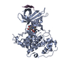
SHEET DETERMINATION METHOD: DSSP THE SHEETS PRESENTED AS "AA" IN EACH CHAIN ON SHEET RECORDS BELOW ... SHEET DETERMINATION METHOD: DSSP THE SHEETS PRESENTED AS "AA" IN EACH CHAIN ON SHEET RECORDS BELOW IS ACTUALLY AN 6-STRANDED BARREL THIS IS REPRESENTED BY A 7-STRANDED SHEET IN WHICH THE FIRST AND LAST STRANDS ARE IDENTICAL. THE SHEETS PRESENTED AS "BA" IN EACH CHAIN ON SHEET RECORDS BELOW IS ACTUALLY AN 5-STRANDED BARREL THIS IS REPRESENTED BY A 6-STRANDED SHEET IN WHICH THE FIRST AND LAST STRANDS ARE IDENTICAL.
Resolution: 2.35→88.74 Å / Cor.coef. Fo:Fc: 0.954 / Cor.coef. Fo:Fc free: 0.938 / SU B: 9.346 / SU ML: 0.112 / Cross valid method: THROUGHOUT / ESU R: 0.214 / ESU R Free: 0.186 / Stereochemistry target values: MAXIMUM LIKELIHOOD / Details: HYDROGENS HAVE BEEN ADDED IN THE RIDING POSITIONS.
Rfactor
Num. reflection
% reflection
Selection details
Rfree
0.227
2638
5.1 %
RANDOM
Rwork
0.19
-
-
-
obs
0.192
49335
96.9 %
-
Solvent computation
Ion probe radii: 0.8 Å / Shrinkage radii: 0.8 Å / VDW probe radii: 1.2 Å / Solvent model: MASK
 Movie
Movie Controller

 Yorodumi
 Open data
 Basic information
 Components
 Keywords
 Function and homology information
 HOMO SAPIENS (human)
 X-RAY DIFFRACTION /
  Authors
 Citation
 Structure visualization
 Downloads & links
 Other downloads


 PDBj
PDBj


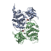
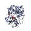
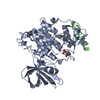
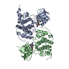
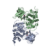
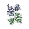
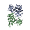
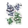
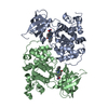
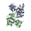
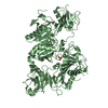
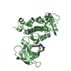
 Assembly
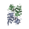

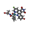
 Mass: 629.515 Da / Num. of mol.: 2 / Source method: obtained synthetically / Formula: C27H17FN4O7Ru
 Mass: 18.015 Da / Num. of mol.: 182 / Source method: isolated from a natural source / Formula: H2O
 Sample preparation
 / Beamline: A1 / Wavelength: 0.978
  Processing