retinal rod cell apoptotic process / PDE3B signalling / cellular response to high light intensity / Inhibition of TSC complex formation by AKT (PKB) / positive regulation of cap-dependent translational initiation / AKT-mediated inactivation of FOXO1A / negative regulation of long-chain fatty acid import across plasma membrane / Negative regulation of the PI3K/AKT network / Activation of AKT2 / AKT phosphorylates targets in the nucleus ...retinal rod cell apoptotic process / PDE3B signalling / cellular response to high light intensity / Inhibition of TSC complex formation by AKT (PKB) / positive regulation of cap-dependent translational initiation / AKT-mediated inactivation of FOXO1A / negative regulation of long-chain fatty acid import across plasma membrane / Negative regulation of the PI3K/AKT network / Activation of AKT2 / AKT phosphorylates targets in the nucleus / negative regulation of glycogen (starch) synthase activity / neuron projection organization / regulation of microtubule anchoring at centrosome / negative regulation of mesenchymal stem cell differentiation / negative regulation of type B pancreatic cell development / superior temporal gyrus development / positive regulation of protein localization to cilium / negative regulation of glycogen biosynthetic process / positive regulation of fatty acid beta-oxidation / positive regulation of glucose metabolic process / negative regulation of TORC2 signaling / RUNX2 regulates genes involved in cell migration / beta-arrestin-dependent dopamine receptor signaling pathway / negative regulation of dopaminergic neuron differentiation / positive regulation of protein localization to centrosome / maintenance of cell polarity / mammary gland epithelial cell differentiation / positive regulation of cilium assembly / CRMPs in Sema3A signaling / tau-protein kinase / heart valve development / beta-catenin destruction complex / RAB GEFs exchange GTP for GDP on RABs / APC truncation mutants have impaired AXIN binding / AXIN missense mutants destabilize the destruction complex / Truncations of AMER1 destabilize the destruction complex / glycogen biosynthetic process / peripheral nervous system myelin maintenance / positive regulation of mitochondrial outer membrane permeabilization involved in apoptotic signaling pathway / regulation of protein export from nucleus / cellular response to interleukin-3 / Maturation of nucleoprotein / Beta-catenin phosphorylation cascade / Signaling by GSK3beta mutants / CTNNB1 S33 mutants aren't phosphorylated / CTNNB1 S37 mutants aren't phosphorylated / CTNNB1 S45 mutants aren't phosphorylated / CTNNB1 T41 mutants aren't phosphorylated / regulation of long-term synaptic potentiation / Wnt signalosome / negative regulation of TOR signaling / regulation of microtubule-based process / AKT phosphorylates targets in the cytosol / Disassembly of the destruction complex and recruitment of AXIN to the membrane / positive regulation of protein binding / regulation of axon extension / negative regulation of calcineurin-NFAT signaling cascade / positive regulation of cell motility / Maturation of nucleoprotein / Regulation of TP53 Activity through Association with Co-factors / negative regulation of protein localization to nucleus / negative regulation of epithelial to mesenchymal transition / tau-protein kinase activity / positive regulation of cell-matrix adhesion / Co-inhibition by CTLA4 / glycogen metabolic process / ER overload response / regulation of axonogenesis / regulation of dendrite morphogenesis / regulation of neuron projection development / negative regulation of PERK-mediated unfolded protein response / Constitutive Signaling by AKT1 E17K in Cancer / protein kinase A catalytic subunit binding / establishment of cell polarity / Regulation of MITF-M-dependent genes involved in pigmentation / Regulation of localization of FOXO transcription factors / dynactin binding / epithelial to mesenchymal transition / CD28 dependent PI3K/Akt signaling / Regulation of HSF1-mediated heat shock response / Activation of BAD and translocation to mitochondria / positive regulation of blood vessel endothelial cell migration / Estrogen-dependent nuclear events downstream of ESR-membrane signaling / positive regulation of glycogen biosynthetic process / positive regulation of protein targeting to membrane / canonical Wnt signaling pathway / SARS-CoV-2 targets host intracellular signalling and regulatory pathways / negative regulation of osteoblast differentiation / fat cell differentiation / NF-kappaB binding / Cyclin E associated events during G1/S transition / negative regulation of extrinsic apoptotic signaling pathway via death domain receptors / Cyclin A:Cdk2-associated events at S phase entry / negative regulation of protein-containing complex assembly / regulation of cellular response to heat / Regulation of TP53 Activity through Acetylation / extrinsic apoptotic signaling pathway / cellular response to retinoic acid / extrinsic apoptotic signaling pathway in absence of ligand / positive regulation of type I interferon production Similarity search - Function
Protein Kinase B beta, catalytic domain / Protein Kinase B, pleckstrin homology domain / Glycogen synthase kinase 3, catalytic domain / : / Protein kinase, C-terminal / Protein kinase C terminal domain / Extension to Ser/Thr-type protein kinases / PH domain / AGC-kinase, C-terminal / AGC-kinase C-terminal domain profile. ...Protein Kinase B beta, catalytic domain / Protein Kinase B, pleckstrin homology domain / Glycogen synthase kinase 3, catalytic domain / : / Protein kinase, C-terminal / Protein kinase C terminal domain / Extension to Ser/Thr-type protein kinases / PH domain / AGC-kinase, C-terminal / AGC-kinase C-terminal domain profile. / PH domain profile. / Pleckstrin homology domain. / Pleckstrin homology domain / PH-like domain superfamily / Phosphorylase Kinase; domain 1 / Phosphorylase Kinase; domain 1 / Transferase(Phosphotransferase) domain 1 / Transferase(Phosphotransferase); domain 1 / Serine/threonine-protein kinase, active site / Serine/Threonine protein kinases active-site signature. / Protein kinase domain / Serine/Threonine protein kinases, catalytic domain / Protein kinase, ATP binding site / Protein kinases ATP-binding region signature. / Protein kinase domain profile. / Protein kinase domain / Protein kinase-like domain superfamily / 2-Layer Sandwich / Orthogonal Bundle / Mainly Alpha / Alpha Beta Similarity search - Domain/homology
Mass: 18.015 Da / Num. of mol.: 323 / Source method: isolated from a natural source / Formula: H2O
Has protein modification
Y
Sequence details
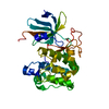
CLONE DOES NOT CONTAIN THE FIRST 145 RESIDUES OF THE P31751 SEQUENCE (PH-DOMAIN). RESIDUES GAMDP AT ...CLONE DOES NOT CONTAIN THE FIRST 145 RESIDUES OF THE P31751 SEQUENCE (PH-DOMAIN). RESIDUES GAMDP AT THE START OF THE CLONED SEQUENCE ARE ARTEFACTS FROM A PURIFICATION TAG, HOWEVER THESE ARE NOT VISIBLE IN THE STRUCTURE AND WERE NOT BUILT. THE CLONE CONTAINS THE SEQUENCE EEQEMFEDFDYIADW (PIFTIDE) INSTEAD OF THE P31751 SEQUENCE FROM POSITION 465 ONWARDS. RESIDUES 450 - 468 ARE DISORDERED AND HAVE NOT BEEN BUILT IN THE STRUCTURE. RESIDUES 3-12 ONLY
-
Experimental details
-
Experiment
Experiment
Method: X-RAY DIFFRACTION
-
Sample preparation
Crystal
Density Matthews: 2.15 Å3/Da / Density % sol: 42.46 %
Method to determine structure: OTHER / Resolution: 1.8→55.19 Å / Cor.coef. Fo:Fc: 0.957 / Cor.coef. Fo:Fc free: 0.942 / SU B: 2.454 / SU ML: 0.077 / Cross valid method: THROUGHOUT / ESU R: 0.13 / ESU R Free: 0.122 / Stereochemistry target values: MAXIMUM LIKELIHOOD / Details: HYDROGENS HAVE BEEN ADDED IN THE RIDING POSITIONS.
Rfactor
Num. reflection
% reflection
Selection details
Rfree
0.21
1676
5.1 %
RANDOM
Rwork
0.175
-
-
-
obs
0.177
31145
94.6 %
-
Solvent computation
Ion probe radii: 0.8 Å / Shrinkage radii: 0.8 Å / VDW probe radii: 1.2 Å / Solvent model: BABINET MODEL WITH MASK
 Movie
Movie Controller

 Yorodumi
 Open data
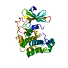
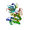
 Basic information
 Components
 Keywords
 Function and homology information
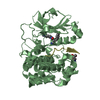
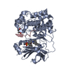
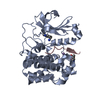
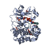
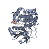
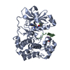
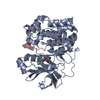
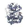
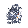
 HOMO SAPIENS (human)
 X-RAY DIFFRACTION /
  Authors
 Citation
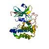
 Structure visualization
 Downloads & links
 Other downloads


 PDBj
PDBj


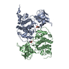
 Assembly
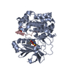

 SPODOPTERA FRUGIPERDA (fall armyworm)
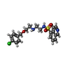
 Mass: 419.925 Da / Num. of mol.: 1 / Source method: obtained synthetically / Formula: C20H22ClN3O3S
 Mass: 62.068 Da / Num. of mol.: 1 / Source method: obtained synthetically / Formula: C2H6O2
 Mass: 18.015 Da / Num. of mol.: 323 / Source method: isolated from a natural source / Formula: H2O
 Sample preparation
 / Beamline: ID29 / Wavelength: 0.9762
  Processing