positive regulation of endodeoxyribonuclease activity / regulation of tRNA methylation / negative regulation of protein maturation / negative regulation of fatty acid beta-oxidation / positive regulation of protein localization to endoplasmic reticulum / regulation of glycogen biosynthetic process / negative regulation of lymphocyte migration / cellular response to decreased oxygen levels / negative regulation of protein localization to lysosome / cellular response to rapamycin ...positive regulation of endodeoxyribonuclease activity / regulation of tRNA methylation / negative regulation of protein maturation / negative regulation of fatty acid beta-oxidation / positive regulation of protein localization to endoplasmic reticulum / regulation of glycogen biosynthetic process / negative regulation of lymphocyte migration / cellular response to decreased oxygen levels / negative regulation of protein localization to lysosome / cellular response to rapamycin / maintenance of protein location in mitochondrion / mammalian oogenesis stage / AKT-mediated inactivation of FOXO1A / negative regulation of long-chain fatty acid import across plasma membrane / Negative regulation of the PI3K/AKT network / regulation of type B pancreatic cell development / maternal placenta development / positive regulation of anaphase-promoting complex-dependent catabolic process / : / potassium channel activator activity / AKT phosphorylates targets in the nucleus / activation-induced cell death of T cells / cellular response to oxidised low-density lipoprotein particle stimulus / negative regulation of cilium assembly / negative regulation of hydrogen peroxide-induced neuron intrinsic apoptotic signaling pathway / Butyrate Response Factor 1 (BRF1) binds and destabilizes mRNA / positive regulation of TORC2 signaling / positive regulation of glucose metabolic process / RUNX2 regulates genes involved in cell migration / beta-arrestin-dependent dopamine receptor signaling pathway / cellular response to peptide / positive regulation of organ growth / mammary gland epithelial cell differentiation / interleukin-18-mediated signaling pathway / fibroblast migration / positive regulation of sodium ion transport / MTOR signalling / response to growth factor / response to fluid shear stress / cellular response to granulocyte macrophage colony-stimulating factor stimulus / negative regulation of leukocyte cell-cell adhesion / RAB GEFs exchange GTP for GDP on RABs / glycogen biosynthetic process / peripheral nervous system myelin maintenance / cell migration involved in sprouting angiogenesis / phosphatidylinositol-3,4-bisphosphate binding / positive regulation of protein localization to cell surface / phosphorylation / complement receptor mediated signaling pathway / sphingosine-1-phosphate receptor signaling pathway / AKT phosphorylates targets in the cytosol / anoikis / regulation of postsynapse organization / positive regulation of fibroblast migration / response to growth hormone / labyrinthine layer blood vessel development / execution phase of apoptosis / regulation of myelination / sperm glycocalyx / Regulation of TP53 Activity through Association with Co-factors / KSRP (KHSRP) binds and destabilizes mRNA / response to UV-A / peptidyl-threonine phosphorylation / TORC2 complex binding / Mechanical load activates signaling by PIEZO1 and integrins in osteocytes / Co-inhibition by CTLA4 / perinuclear theca / negative regulation of cGAS/STING signaling pathway / negative regulation of macroautophagy / negative regulation of release of cytochrome c from mitochondria / cellular response to stress / response to food / negative regulation of PERK-mediated unfolded protein response / regulation of neuron projection development / Constitutive Signaling by AKT1 E17K in Cancer / apoptotic mitochondrial changes / phosphatidylinositol-3,4,5-trisphosphate binding / positive regulation of protein metabolic process / Regulation of localization of FOXO transcription factors / behavioral response to pain / cellular response to vascular endothelial growth factor stimulus / TOR signaling / positive regulation of peptidyl-serine phosphorylation / negative regulation of Notch signaling pathway / CD28 dependent PI3K/Akt signaling / Activation of BAD and translocation to mitochondria / Estrogen-dependent nuclear events downstream of ESR-membrane signaling / positive regulation of blood vessel endothelial cell migration / positive regulation of glycogen biosynthetic process / negative regulation of extrinsic apoptotic signaling pathway in absence of ligand / Mitochondrial unfolded protein response (UPRmt) / protein serine/threonine kinase inhibitor activity / response to insulin-like growth factor stimulus / SARS-CoV-2 targets host intracellular signalling and regulatory pathways / positive regulation of fat cell differentiation / positive regulation of lipid biosynthetic process / negative regulation of oxidative stress-induced intrinsic apoptotic signaling pathway / Cyclin E associated events during G1/S transition / positive regulation of G1/S transition of mitotic cell cycle / Cyclin A:Cdk2-associated events at S phase entry Similarity search - Function
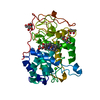
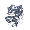
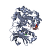
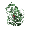
Protein kinase B alpha, catalytic domain / Protein Kinase B, pleckstrin homology domain / Protein kinase, C-terminal / Protein kinase C terminal domain / Extension to Ser/Thr-type protein kinases / AGC-kinase, C-terminal / PH domain / AGC-kinase C-terminal domain profile. / PH domain profile. / Pleckstrin homology domain. ...Protein kinase B alpha, catalytic domain / Protein Kinase B, pleckstrin homology domain / Protein kinase, C-terminal / Protein kinase C terminal domain / Extension to Ser/Thr-type protein kinases / AGC-kinase, C-terminal / PH domain / AGC-kinase C-terminal domain profile. / PH domain profile. / Pleckstrin homology domain. / Pleckstrin homology domain / PH-like domain superfamily / Phosphorylase Kinase; domain 1 / Phosphorylase Kinase; domain 1 / Transferase(Phosphotransferase) domain 1 / Transferase(Phosphotransferase); domain 1 / Serine/threonine-protein kinase, active site / Serine/Threonine protein kinases active-site signature. / Protein kinase domain / Serine/Threonine protein kinases, catalytic domain / Protein kinase, ATP binding site / Protein kinases ATP-binding region signature. / Protein kinase domain profile. / Protein kinase domain / Protein kinase-like domain superfamily / 2-Layer Sandwich / Orthogonal Bundle / Mainly Alpha / Alpha Beta Similarity search - Domain/homology
In the structure databanks used in Yorodumi, some data are registered as the other names, "COVID-19 virus" and "2019-nCoV". Here are the details of the virus and the list of structure data.
Jan 31, 2019. EMDB accession codes are about to change! (news from PDBe EMDB page)
EMDB accession codes are about to change! (news from PDBe EMDB page)
The allocation of 4 digits for EMDB accession codes will soon come to an end. Whilst these codes will remain in use, new EMDB accession codes will include an additional digit and will expand incrementally as the available range of codes is exhausted. The current 4-digit format prefixed with “EMD-” (i.e. EMD-XXXX) will advance to a 5-digit format (i.e. EMD-XXXXX), and so on. It is currently estimated that the 4-digit codes will be depleted around Spring 2019, at which point the 5-digit format will come into force.
The EM Navigator/Yorodumi systems omit the EMD- prefix.
Related info.:Q: What is EMD? / ID/Accession-code notation in Yorodumi/EM Navigator
Yorodumi is a browser for structure data from EMDB, PDB, SASBDB, etc.
This page is also the successor to EM Navigator detail page, and also detail information page/front-end page for Omokage search.
The word "yorodu" (or yorozu) is an old Japanese word meaning "ten thousand". "mi" (miru) is to see.
Related info.:EMDB / PDB / SASBDB / Comparison of 3 databanks / Yorodumi Search / Aug 31, 2016. New EM Navigator & Yorodumi / Yorodumi Papers / Jmol/JSmol / Function and homology information / Changes in new EM Navigator and Yorodumi
 Movie
Movie Controller


 Open data
 Basic information
 Components
 Keywords
 Function and homology information
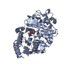
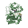
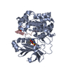
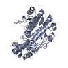
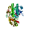
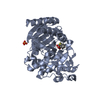
 Homo sapiens (human)
 X-RAY DIFFRACTION /
  Authors
 Citation
 Structure visualization
 Downloads & links
 Other downloads
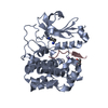


 PDBj
PDBj


 Assembly
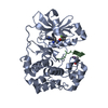
 unidentified baculovirus / Strain (production host): Sf9
 Mass: 54.938 Da / Num. of mol.: 1 / Source method: obtained synthetically / Formula: Mn
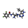
 Mass: 400.425 Da / Num. of mol.: 1 / Source method: obtained synthetically / Formula: C20H22F2N6O
 Mass: 18.015 Da / Num. of mol.: 247 / Source method: isolated from a natural source / Formula: H2O
 Sample preparation
 Processing